Summary information and primary citation
- PDB-id
- 5mht; SNAP-derived features in text and JSON formats;
DNAproDB
- Class
- transferase-DNA
- Method
- X-ray (2.7 Å)
- Summary
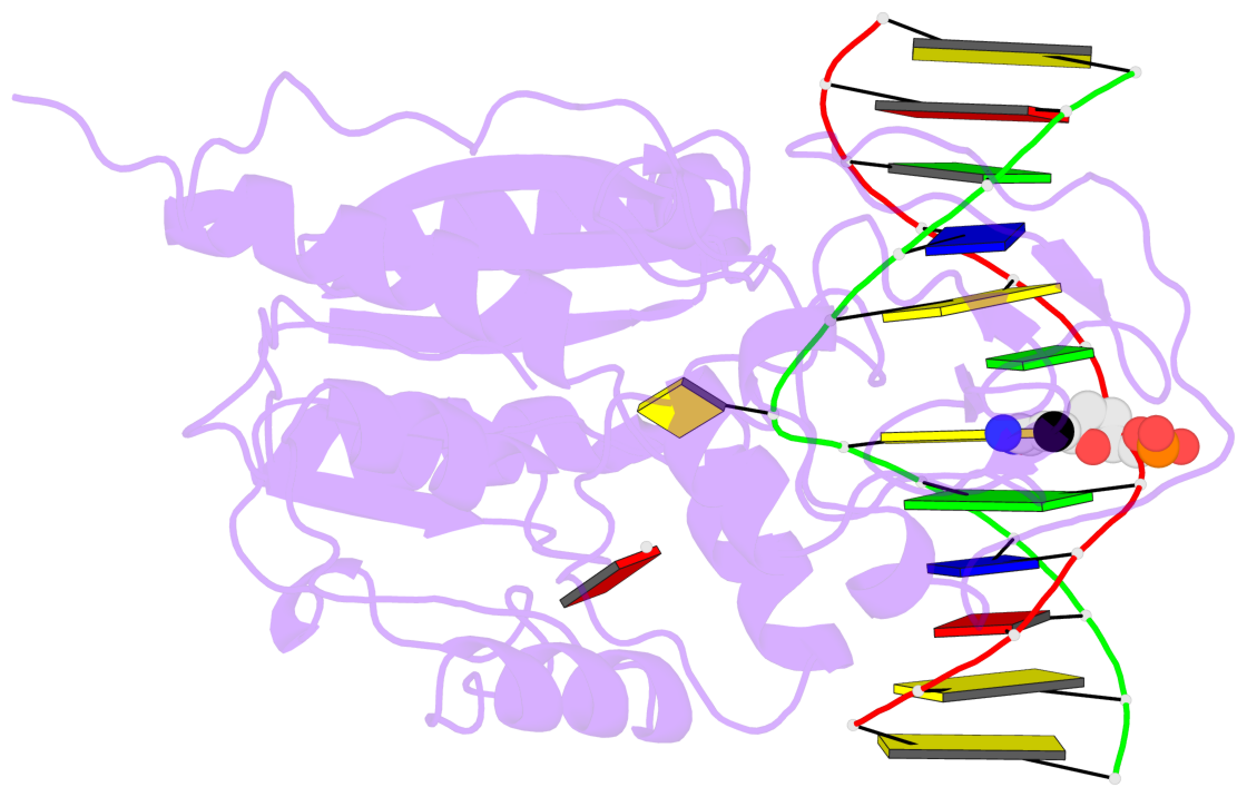
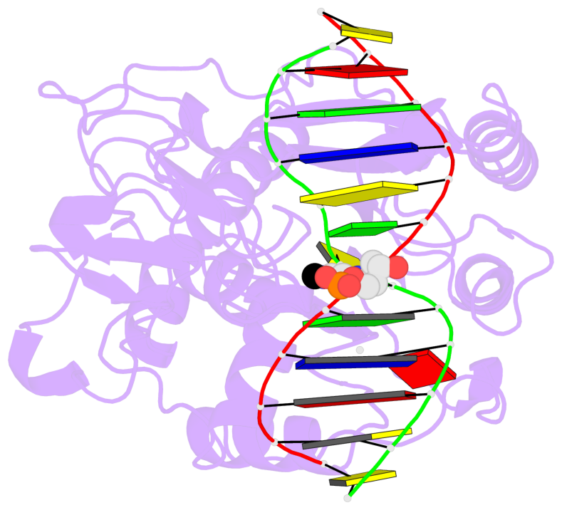
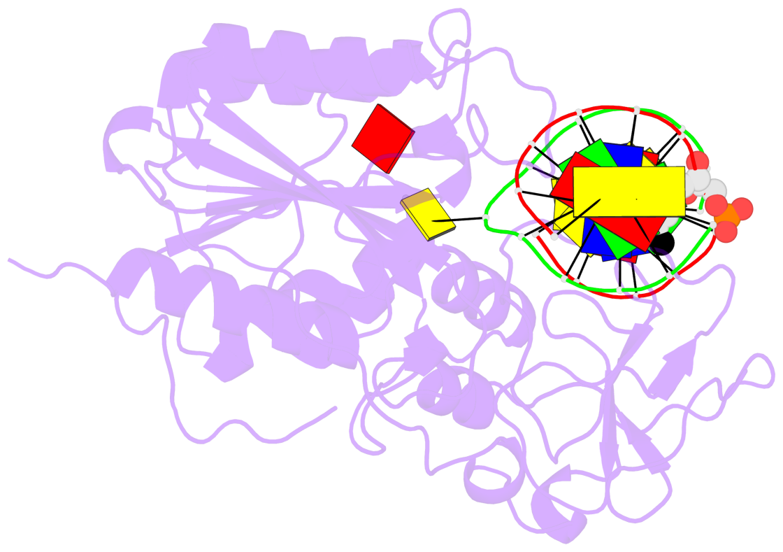
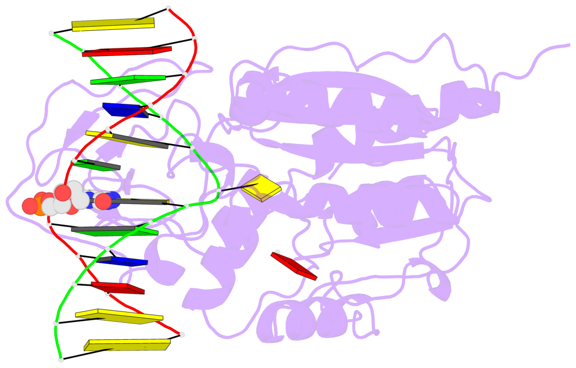
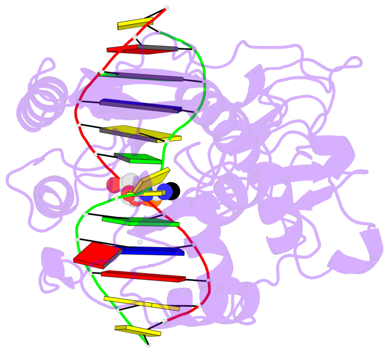
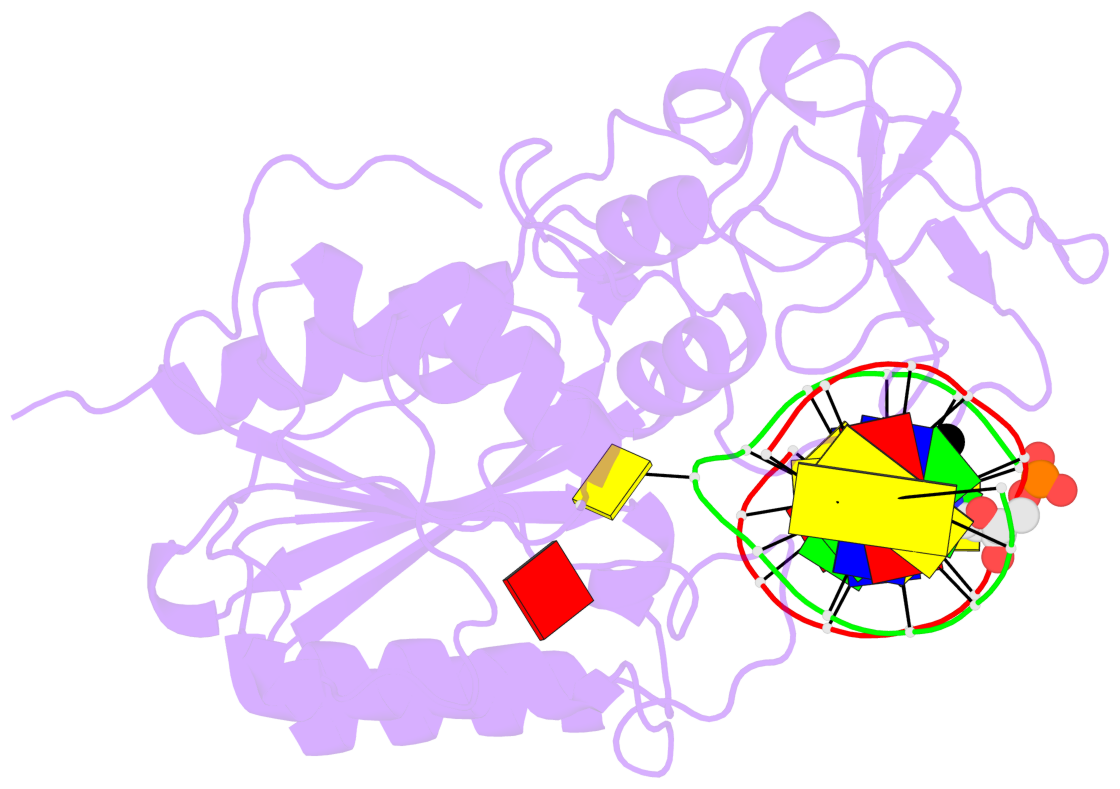
- Ternary structure of hhai methyltransferase with hemimethylated DNA and adohcy
- Reference
- O'Gara M, Roberts RJ, Cheng X (1996): "A structural basis for the preferential binding of hemimethylated DNA by HhaI DNA methyltransferase." J.Mol.Biol., 263, 597-606. doi: 10.1006/jmbi.1996.0601.
- Abstract
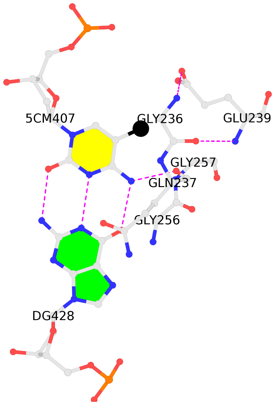
- The crystal structure of HhaI methyltransferase complexed with non-palindromic duplex DNA, containing a hemimethylated recognition sequence, and with the cofactor analog S-adenosyl-L-homocysteine (AdoHcy), has been determined. The structure provides an explanation for the stronger affinities of DNA methyltransferases for hemimethylated DNA than for unmethylated or fully methylated DNA in the presence of AdoHcy. The unmethylated target 2'-deoxycytidine flips out of the DNA helix and the CH group at position 5 makes van der Waals' contacts with the sulfur atom of AdoHcy. Selectivity/preference for hemimethylated over fully methylated DNA may thus reflect interactions among the chemical substituent (H or CH3) at the C5 position of the flipped cytosine, protein and the bound AdoHcy. The 5-methyl-2'-deoxycytidine on the complementary strand remains in the DNA helix, with the methyl group almost perpendicular to the carboxylate group of Glu239, which is part of the sequence recognition loop. Thus, selectivity/preference for hemimethylated over unmethylated DNA appears to result largely from van der Waals' contacts between the planar Glu239 carboxylate and the methyl group of the 5-methyl-2'-deoxycytidine. Furthermore, the positive electrostatic potential originating from the bound AdoHcy extends to the DNA phosphate groups flanking the flipped cytosine. The increased binding to DNA by long-range electrostatic interactions should also occur with the methyl donor S-adenosyl-L-methionine.
- The contacts include paired nucleotides (mostly a G in Watson-Crick G-C pairing), and
amino-acids within a 4.5-A distance cutoff to base atoms of 5mC.
- The structure is oriented in the base reference frame of 5mC, allowing for easy comparison
and direct superimposition between entries.
- The black sphere (•) denotes the 5-methyl carbon atom in 5mC.
No. 1 C.5CM407: hydrophobic-with-A.GLY236 hydrophobic-with-A.GLY256 hydrophobic-with-A.GLY257 is-WC-paired is-in-duplex [+]:GcC/GGC |
 |
|