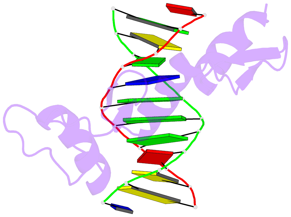
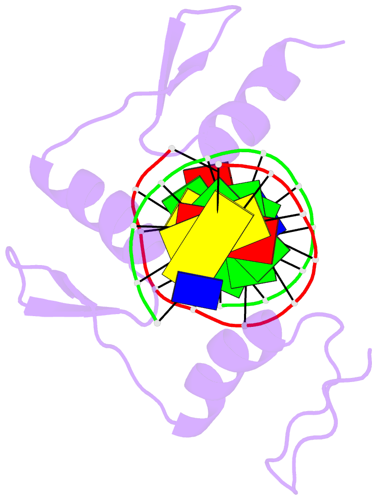
Summary information and primary citation
- PDB-id
- 1a1k; SNAP-derived features in text and JSON formats;
DNAproDB
- Class
- transcription-DNA
- Method
- X-ray (1.9 Å)
- Summary
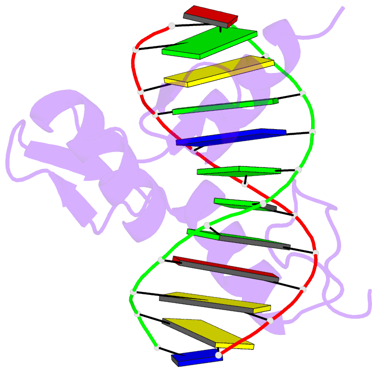
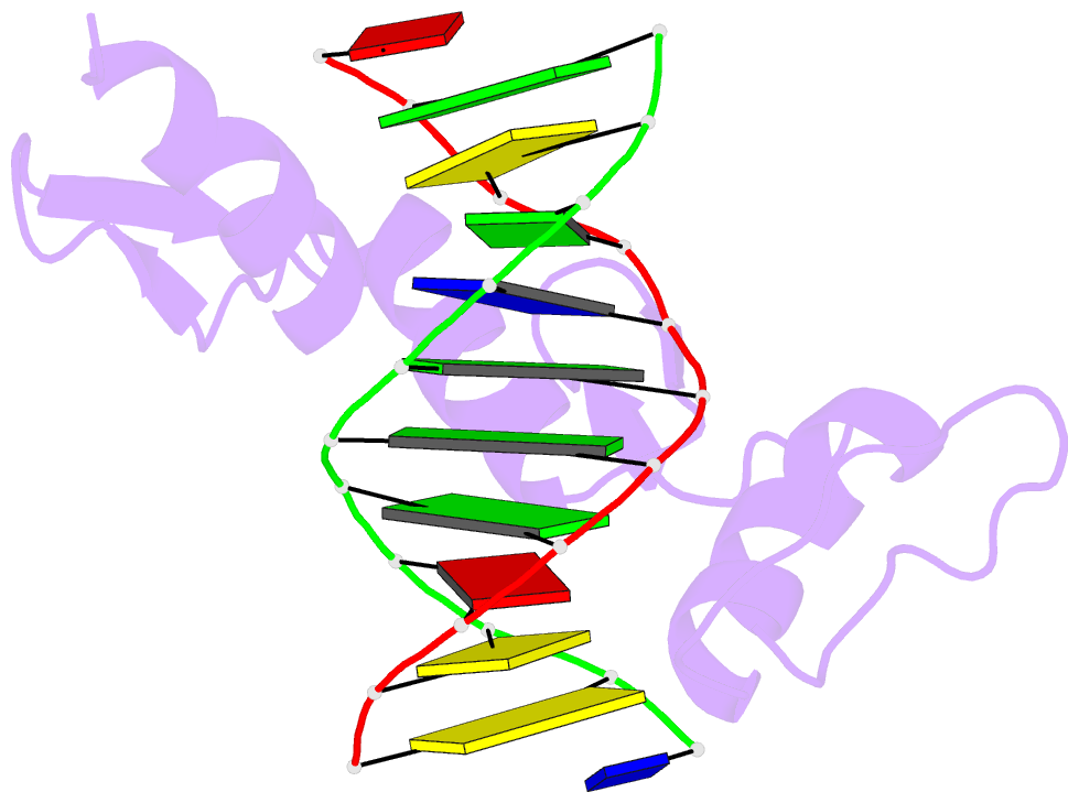
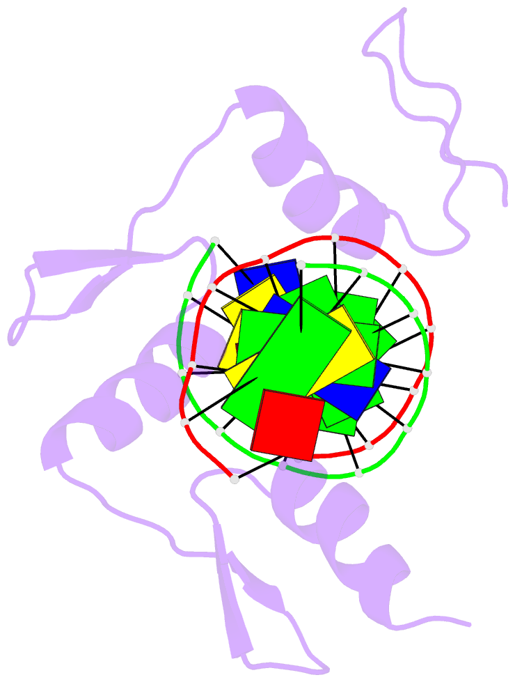
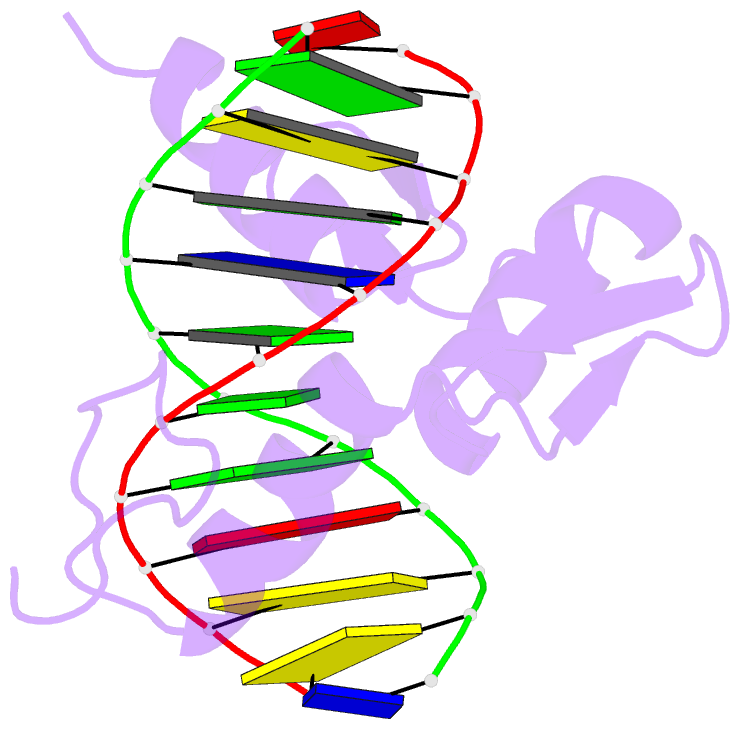
- Radr (zif268 variant) zinc finger-DNA complex (gacc site)
- Reference
- Elrod-Erickson M, Benson TE, Pabo CO (1998): "High-resolution structures of variant Zif268-DNA complexes: implications for understanding zinc finger-DNA recognition." Structure, 6, 451-464. doi: 10.1016/S0969-2126(98)00047-1.
- Abstract
- Background: Zinc fingers of the Cys2-His2 class comprise one of the largest families of eukaryotic DNA-binding motifs and recognize a diverse set of DNA sequences. These proteins have a relatively simple modular structure and key base contacts are typically made by a few residues from each finger. These features make the zinc finger motif an attractive system for designing novel DNA-binding proteins and for exploring fundamental principles of protein-DNA recognition.
Results: Here we report the X-ray crystal structures of zinc finger-DNA complexes involving three variants of Zif268, with multiple changes in the recognition helix of finger one. We describe the structure of each of these three-finger peptides bound to its corresponding target site. To help elucidate the differential basis for site-specific recognition, the structures of four other complexes containing various combinations of these peptides with alternative binding sites have also been determined.
Conclusions: The protein-DNA contacts observed in these complexes reveal the basis for the specificity demonstrated by these Zif268 variants. Many, but not all, of the contacts can be rationalized in terms of a recognition code, but the predictive value of such a code is limited. The structures illustrate how modest changes in the docking arrangement accommodate the new sidechain-base and sidechain-phosphate interactions. Such adaptations help explain the versatility of naturally occurring zinc finger proteins and their utility in design.