Summary information and primary citation
- PDB-id
- 1a34; SNAP-derived features in text and JSON formats;
DNAproDB
- Class
- virus-RNA
- Method
- X-ray (1.81 Å)
- Summary
- Satellite tobacco mosaic virus-RNA complex
- Reference
- Larson SB, Day J, Greenwood A, McPherson A (1998): "Refined structure of satellite tobacco mosaic virus at 1.8 A resolution." J.Mol.Biol., 277, 37-59. doi: 10.1006/jmbi.1997.1570.
- Abstract
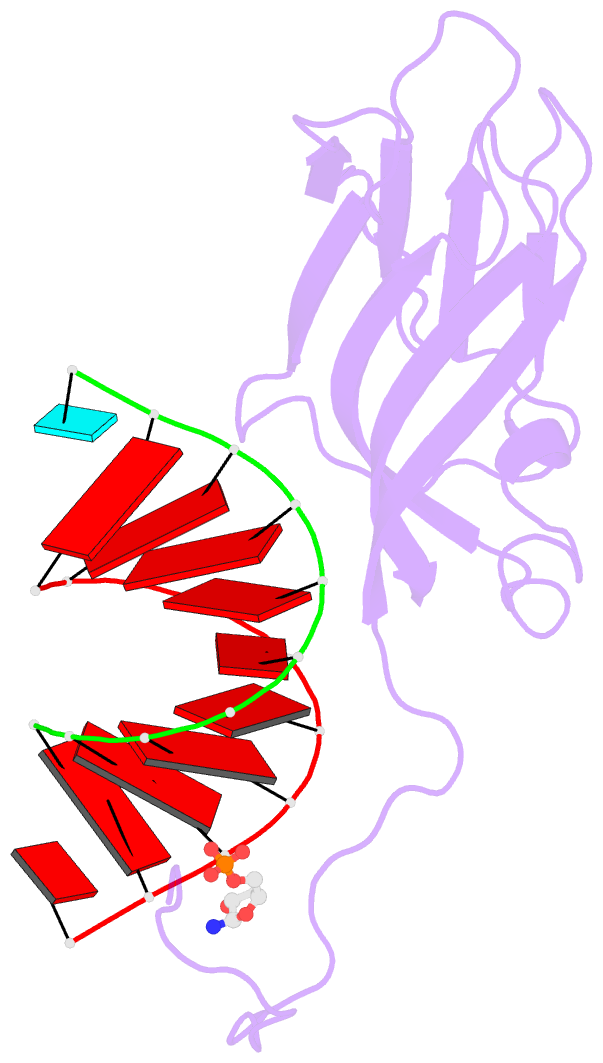
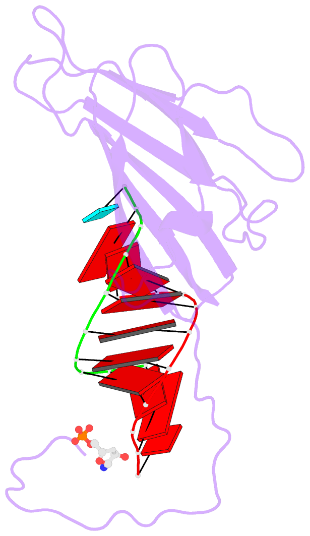
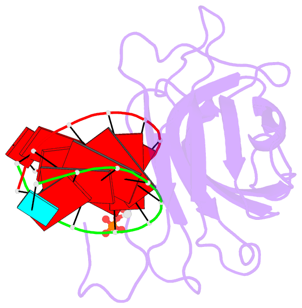
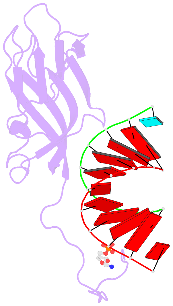
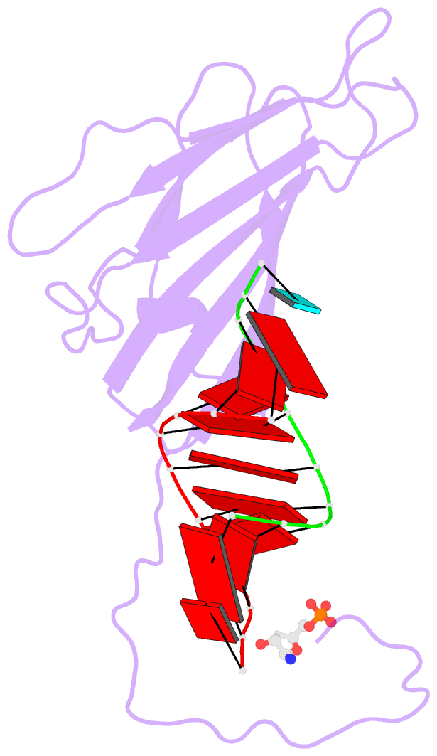
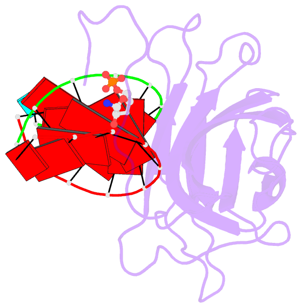
- The molecular structure of satellite tobacco mosaic virus (STMV) has been refined to 1.8 A resolution using X-ray diffraction data collected from crystals grown in microgravity. The final R value was 0.179 and Rfree was 0.184 for 219,086 independent reflections. The final model of the asymmetric unit contained amino acid residues 13 to 159 of a coat protein monomer, 21 nucleotides, a sulfate ion, and 168 water molecules. The nucleotides were visualized as 30 helical segments of nine base-pairs with an additional base stacked at each 3' end, plus a "free" nucleotide, not belonging to the helical segments, but firmly bound by the protein. Sulfate ions are located exactly on 5-fold axes and each is coordinated by ten asparagine side-chains. Of the 10,080 structural waters, 168 per asymmetric unit, about 20% serve to bridge the macromolecular components at protein-protein and protein-nucleic acid interfaces. Binding of RNA to the protein involves some salt linkages, particularly to the phosphate of the free nucleotide, but the major contribution is from an intricate network of hydrogen bonds. There are numerous water molecules in the RNA-protein interface, many serving as intermediate hydrogen bond bridges. The sugar-phosphate backbone contributes most of the donors and acceptors for the RNA. The helical RNA conformation is nearest that of A form DNA. The central region of a helical segment is most extensively involved in contacts with protein, and exhibits low thermal parameters which increase dramatically toward the ends. The visible RNA represents approximately 59% of the total nucleic acid in the virion and is derived from the single-stranded genome, which has folded upon itself to form helical segments. Linking of the helices and the free nucleotides in a contiguous and efficient manner severely restricts the disposition of the remaining, unseen nucleic acid. Using the remaining nucleotides it is possible to fold the RNA according to motifs that provide a periodic distribution of RNA structural elements compatible with the icosahedrally symmetrical arrangement seen in the crystallographic structure. The intimate relationship between protein and nucleic acid in STMV suggests an assembly pathway based on the cooperative and coordinated co-condensation of RNA with capsid protein dimers.