Summary information and primary citation
- PDB-id
- 1aoi; SNAP-derived features in text and JSON formats;
DNAproDB
- Class
- DNA binding protein-DNA
- Method
- X-ray (2.8 Å)
- Summary
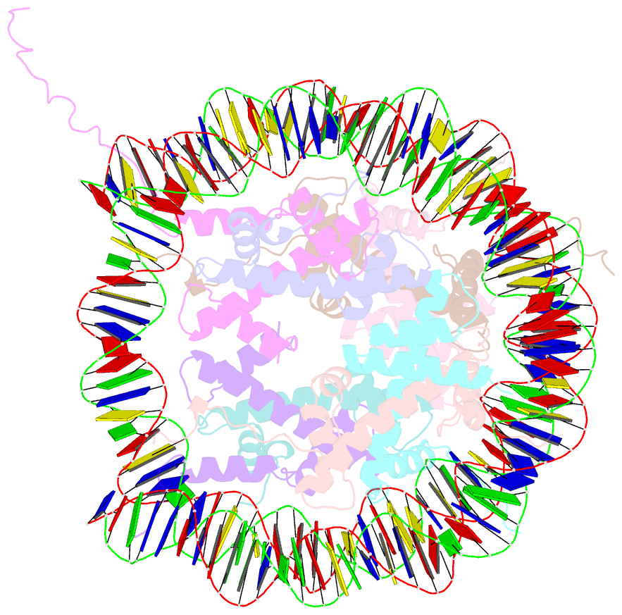
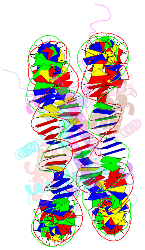
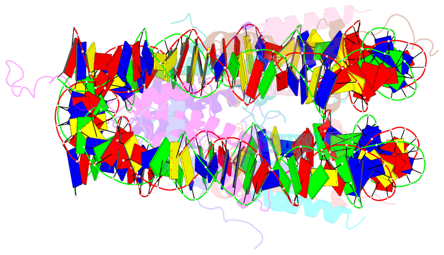
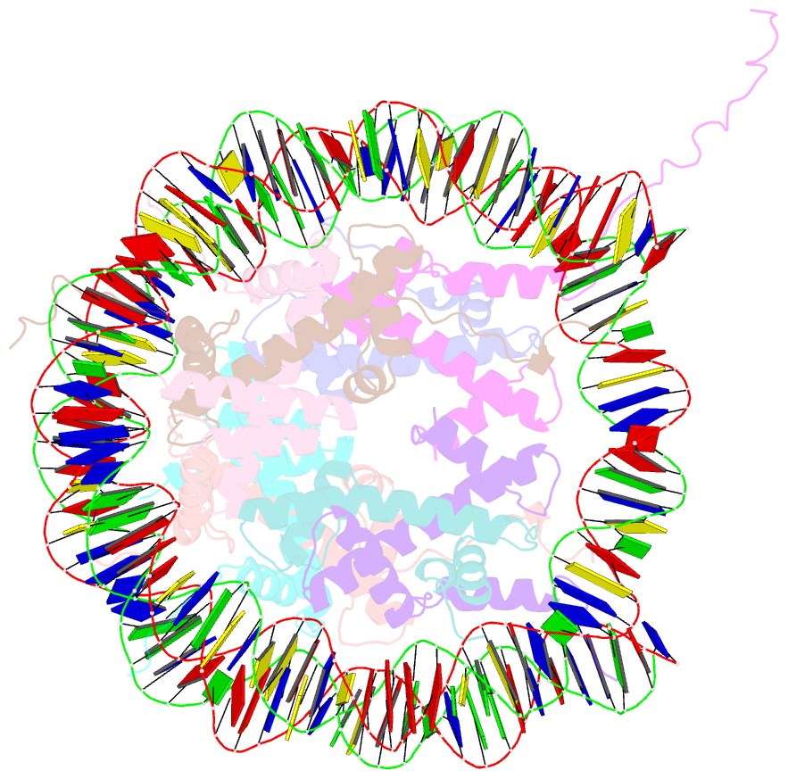
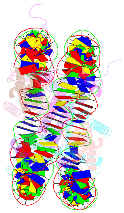
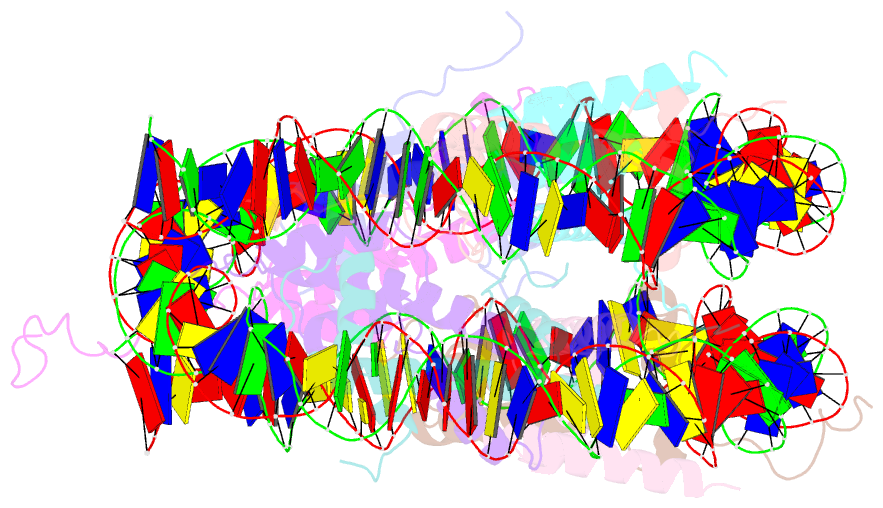
- Complex between nucleosome core particle (h3,h4,h2a,h2b) and 146 bp long DNA fragment
- Reference
- Luger K, Mader AW, Richmond RK, Sargent DF, Richmond TJ (1997): "Crystal structure of the nucleosome core particle at 2.8 A resolution." Nature, 389, 251-260. doi: 10.1038/38444.
- Abstract
- The X-ray crystal structure of the nucleosome core particle of chromatin shows in atomic detail how the histone protein octamer is assembled and how 146 base pairs of DNA are organized into a superhelix around it. Both histone/histone and histone/DNA interactions depend on the histone fold domains and additional, well ordered structure elements extending from this motif. Histone amino-terminal tails pass over and between the gyres of the DNA superhelix to contact neighbouring particles. The lack of uniformity between multiple histone/DNA-binding sites causes the DNA to deviate from ideal superhelix geometry.