Summary information and primary citation
- PDB-id
- 1asy; SNAP-derived features in text and JSON formats;
DNAproDB
- Class
- complex (aminoacyl-trna synthase-trna)
- Method
- X-ray (2.9 Å)
- Summary
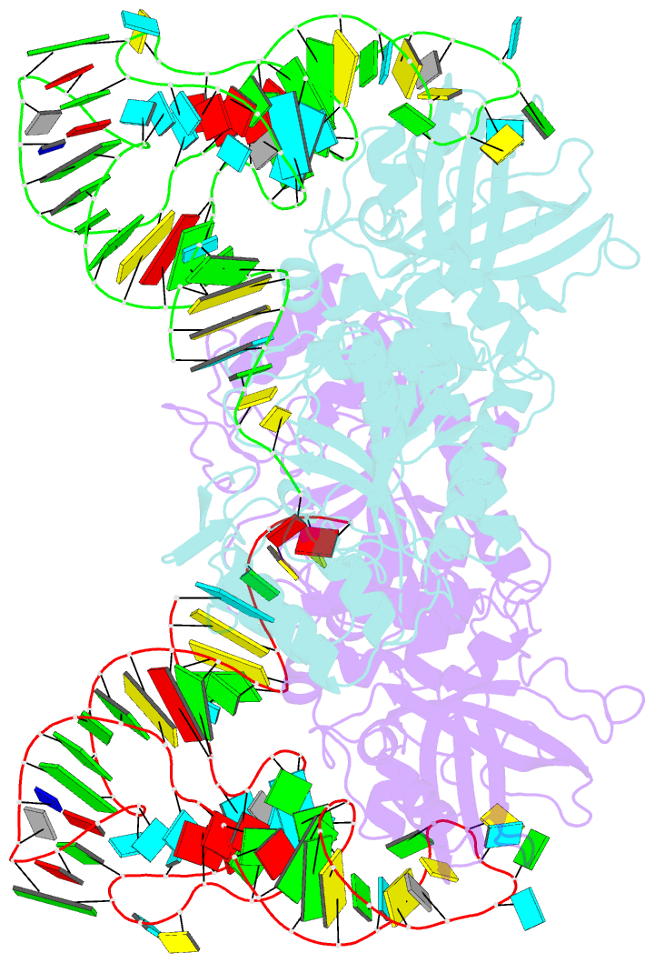
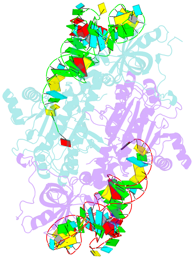
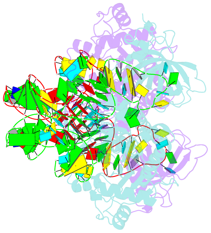
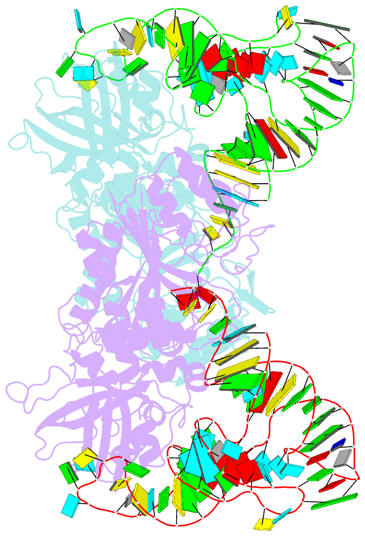
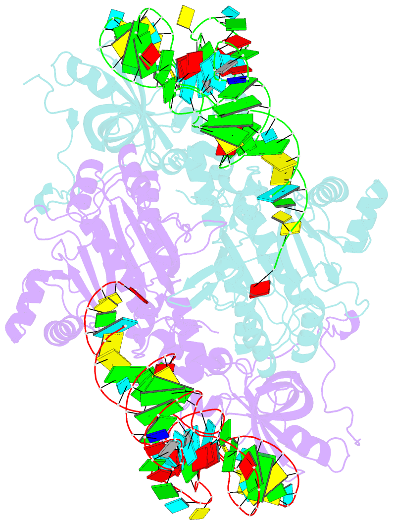
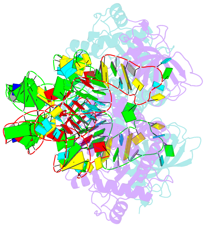
- Class ii aminoacyl transfer RNA synthetases: crystal structure of yeast aspartyl-trna synthetase complexed with trna asp
- Reference
- Ruff M, Krishnaswamy S, Boeglin M, Poterszman A, Mitschler A, Podjarny A, Rees B, Thierry JC, Moras D (1991): "Class II aminoacyl transfer RNA synthetases: crystal structure of yeast aspartyl-tRNA synthetase complexed with tRNA(Asp)." Science, 252, 1682-1689.
- Abstract
- The crystal structure of the binary complex tRNA(Asp)-aspartyl tRNA synthetase from yeast was solved with the use of multiple isomorphous replacement to 3 angstrom resolution. The dimeric synthetase, a member of class II aminoacyl tRNA synthetases (aaRS's) exhibits the characteristic signature motifs conserved in eight aaRS's. These three sequence motifs are contained in the catalytic site domain, built around an antiparallel beta sheet, and flanked by three alpha helices that form the pocket in which adenosine triphosphate (ATP) and the CCA end of tRNA bind. The tRNA(Asp) molecule approaches the synthetase from the variable loop side. The two major contact areas are with the acceptor end and the anticodon stem and loop. In both sites the protein interacts with the tRNA from the major groove side. The correlation between aaRS class II and the initial site of aminoacylation at 3'-OH can be explained by the structure. The molecular association leads to the following features: (i) the backbone of the GCCA single-stranded portion of the acceptor end exhibits a regular helical conformation; (ii) the loop between residues 320 and 342 in motif 2 interacts with the acceptor stem in the major groove and is in contact with the discriminator base G and the first base pair UA; and (iii) the anticodon loop undergoes a large conformational change in order to bind the protein. The conformation of the tRNA molecule in the complex is dictated more by the interaction with the protein than by its own sequence.