Summary information and primary citation
- PDB-id
- 1b2m; SNAP-derived features in text and JSON formats;
DNAproDB
- Class
- hydrolase-RNA
- Method
- X-ray (2.0 Å)
- Summary
- Three-dimensional structure of ribonulcease t1 complexed with an isosteric phosphonate analogue of gpu: alternate substrate binding modes and catalysis.
- Reference
- Arni RK, Watanabe L, Ward RJ, Kreitman RJ, Kumar K, Walz Jr FG (1999): "Three-dimensional structure of ribonuclease T1 complexed with an isosteric phosphonate substrate analogue of GpU: alternate substrate binding modes and catalysis." Biochemistry, 38, 2452-2461. doi: 10.1021/bi982612q.
- Abstract
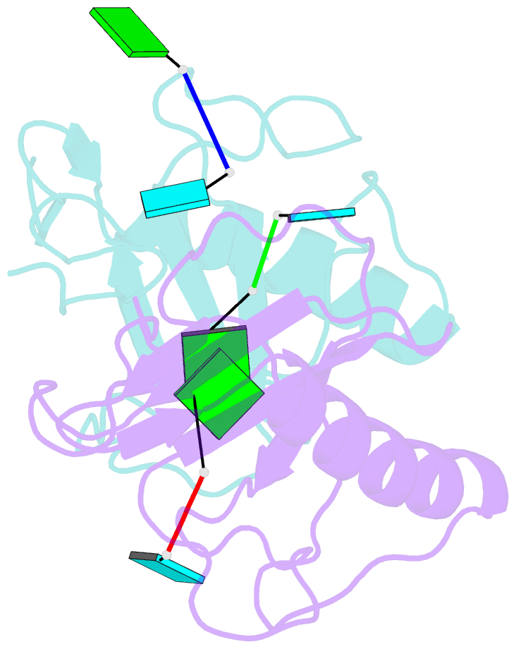
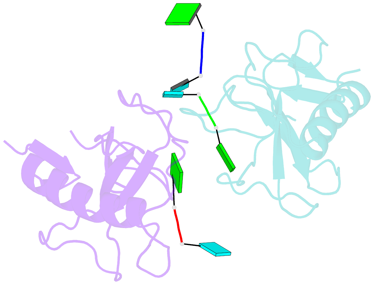
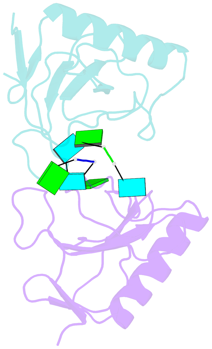
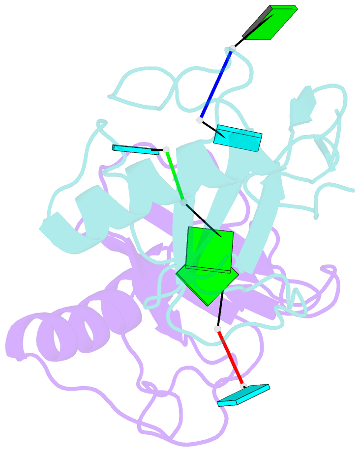
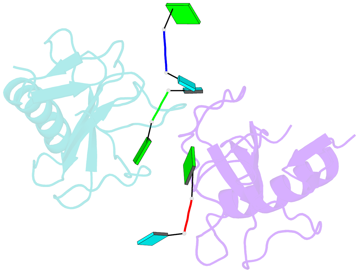
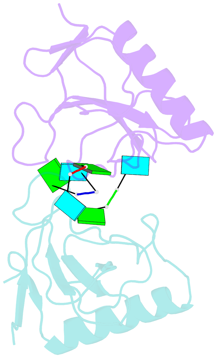
- The X-ray crystal structure of a complex between ribonuclease T1 and guanylyl(3'-6')-6'-deoxyhomouridine (GpcU) has been determined at 2. 0 A resolution. This ligand is an isosteric analogue of the minimal RNA substrate, guanylyl(3'-5')uridine (GpU), where a methylene is substituted for the uridine 5'-oxygen atom. Two protein molecules are part of the asymmetric unit and both have a GpcU bound at the active site in the same manner. The protein-protein interface reveals an extended aromatic stack involving both guanines and three enzyme phenolic groups. A third GpcU has its guanine moiety stacked on His92 at the active site on enzyme molecule A and interacts with GpcU on molecule B in a neighboring unit via hydrogen bonding between uridine ribose 2'- and 3'-OH groups. None of the uridine moieties of the three GpcU molecules in the asymmetric unit interacts directly with the protein. GpcU-active-site interactions involve extensive hydrogen bonding of the guanine moiety at the primary recognition site and of the guanosine 2'-hydroxyl group with His40 and Glu58. On the other hand, the phosphonate group is weakly bound only by a single hydrogen bond with Tyr38, unlike ligand phosphate groups of other substrate analogues and 3'-GMP, which hydrogen-bonded with three additional active-site residues. Hydrogen bonding of the guanylyl 2'-OH group and the phosphonate moiety is essentially the same as that recently observed for a novel structure of a RNase T1-3'-GMP complex obtained immediately after in situ hydrolysis of exo-(Sp)-guanosine 2',3'-cyclophosphorothioate [Zegers et al. (1998) Nature Struct. Biol. 5, 280-283]. It is likely that GpcU at the active site represents a nonproductive binding mode for GpU [Steyaert, J., and Engleborghs (1995) Eur. J. Biochem. 233, 140-144]. The results suggest that the active site of ribonuclease T1 is adapted for optimal tight binding of both the guanylyl 2'-OH and phosphate groups (of GpU) only in the transition state for catalytic transesterification, which is stabilized by adjacent binding of the leaving nucleoside (U) group.