Summary information and primary citation
- PDB-id
- 1b8i; SNAP-derived features in text and JSON formats;
DNAproDB
- Class
- transcription-DNA
- Method
- X-ray (2.4 Å)
- Summary
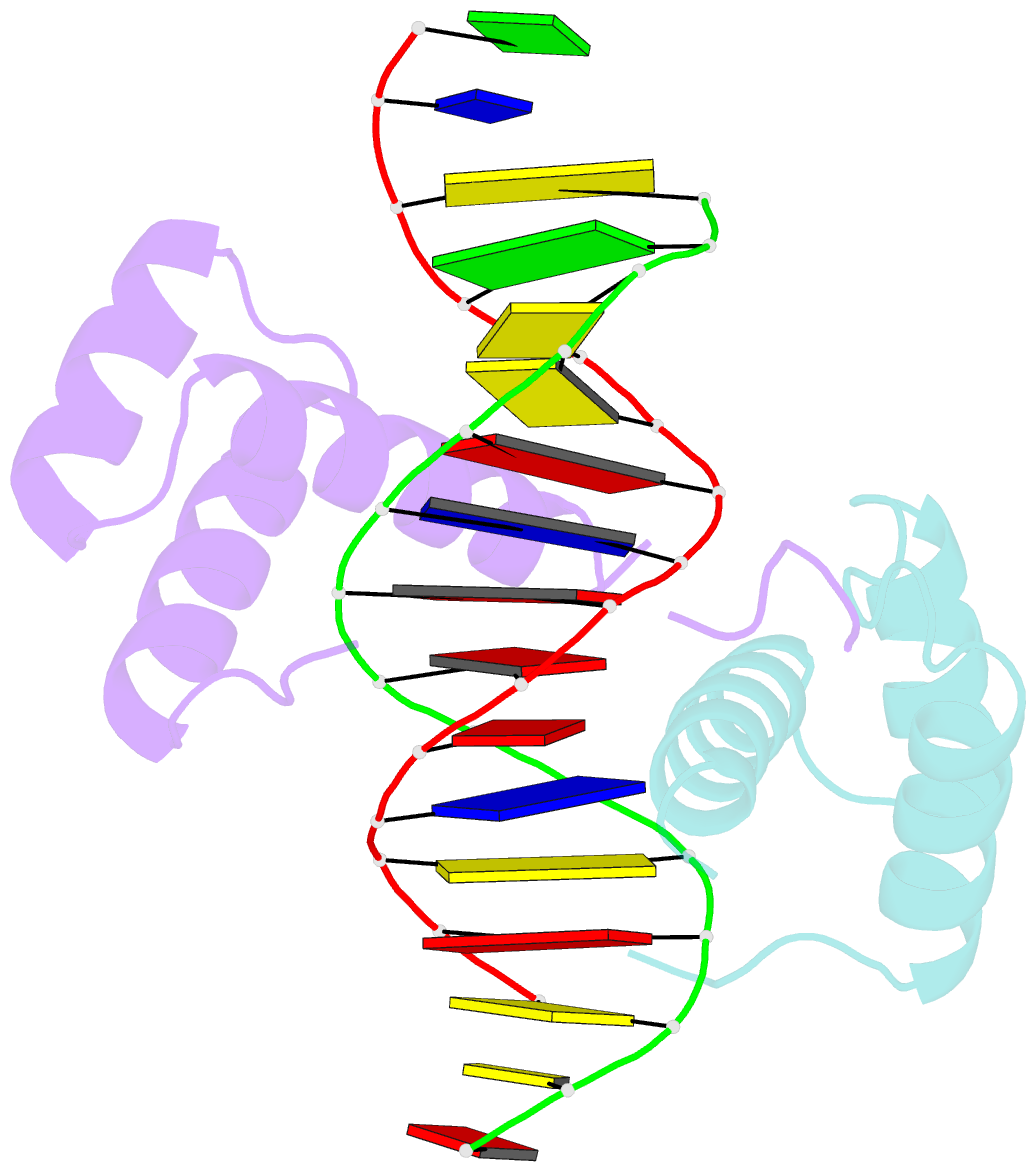
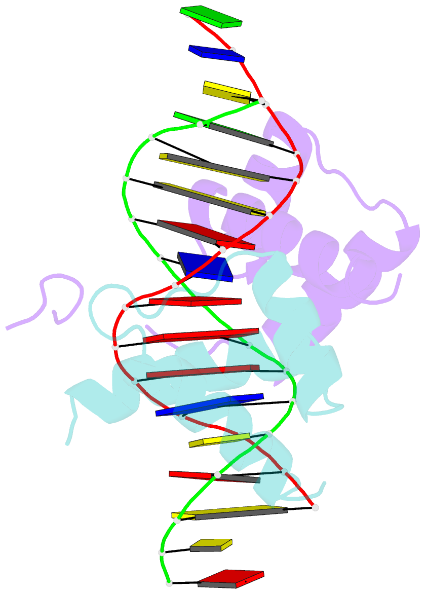
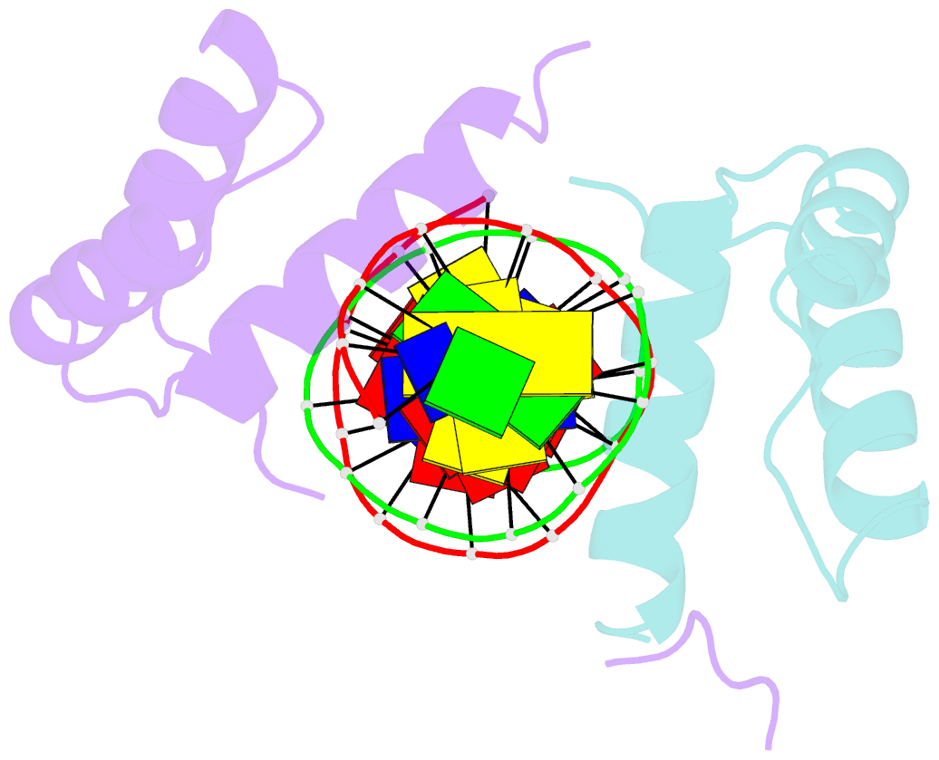
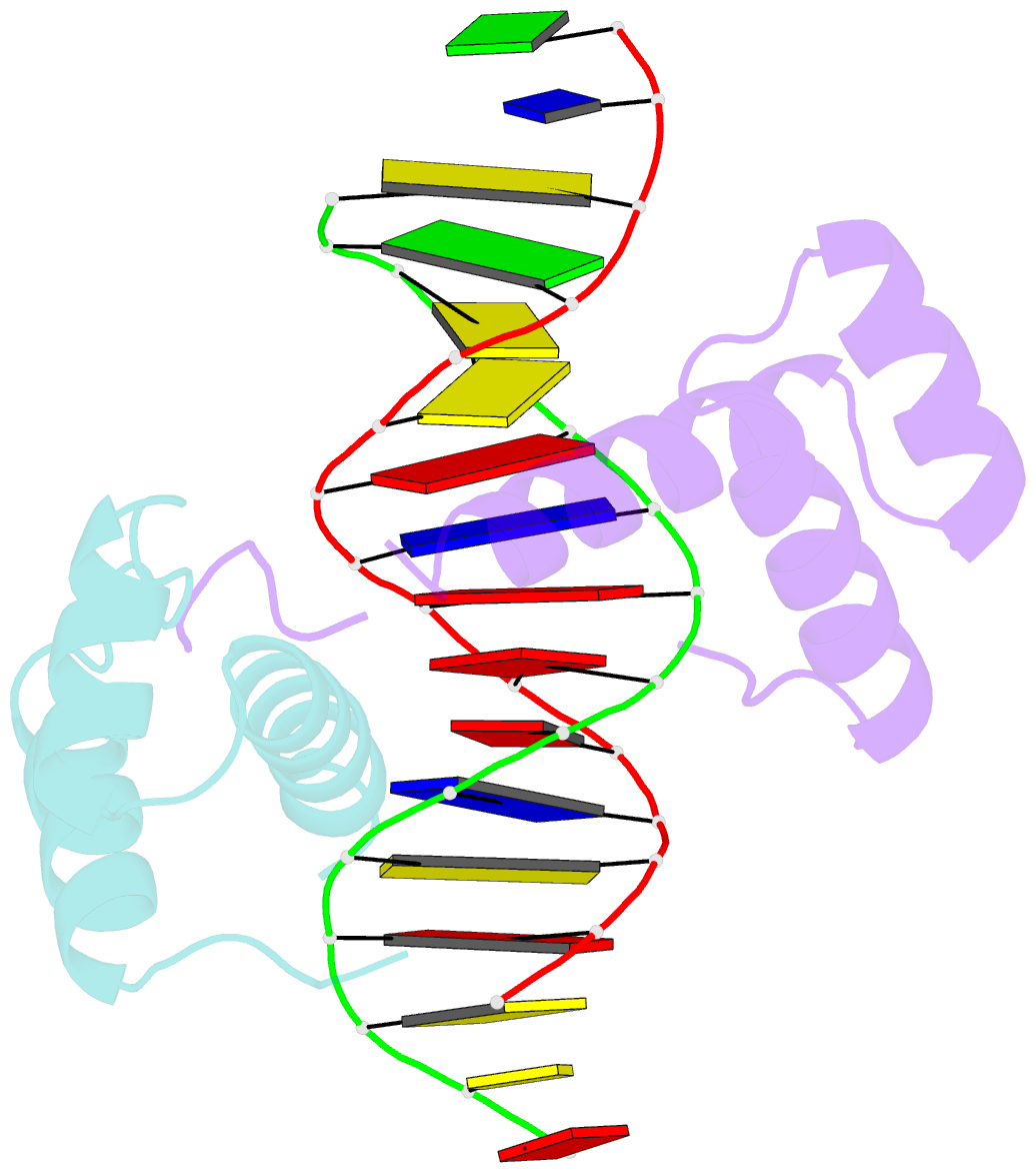
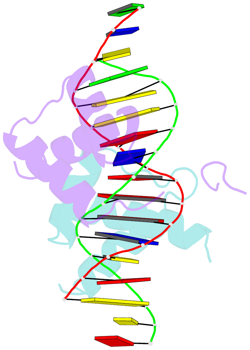
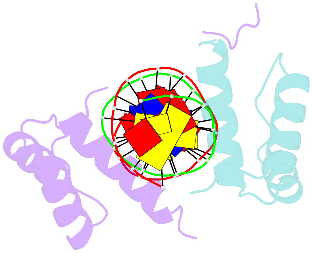
- Structure of the homeotic ubx-exd-DNA ternary complex
- Reference
- Passner JM, Ryoo HD, Shen L, Mann RS, Aggarwal AK (1999): "Structure of a DNA-bound Ultrabithorax-Extradenticle homeodomain complex." Nature, 397, 714-719. doi: 10.1038/17833.
- Abstract
- During the development of multicellular organisms, gene expression must be tightly regulated, both spatially and temporally. One set of transcription factors that are important in animal development is encoded by the homeotic (Hox) genes, which govern the choice between alternative developmental pathways along the anterior-posterior axis. Hox proteins, such as Drosophila Ultrabithorax, have low DNA-binding specificity by themselves but gain affinity and specificity when they bind together with the homeoprotein Extradenticle (or Pbxl in mammals). To understand the structural basis of Hox-Extradenticle pairing, we determine here the crystal structure of an Ultrabithorax-Extradenticle-DNA complex at 2.4 A resolution, using the minimal polypeptides that form a cooperative heterodimer. The Ultrabithorax and Extradenticle homeodomains bind opposite faces of the DNA, with their DNA-recognition helices almost touching each other. However, most of the cooperative interactions arise from the YPWM amino-acid motif of Ultrabithorax-located amino-terminally to its homeodomain-which forms a reverse turn and inserts into a hydrophobic pocket on the Extradenticle homeodomain surface. Together, these protein-DNA and protein-protein interactions define the general principles by which homeotic proteins interact with Extradenticle (or Pbx1) to affect development along the anterior-posterior axis of animals.