Summary information and primary citation
- PDB-id
- 1c04; SNAP-derived features in text and JSON formats;
DNAproDB
- Class
- ribosome
- Method
- X-ray (5.0 Å)
- Summary
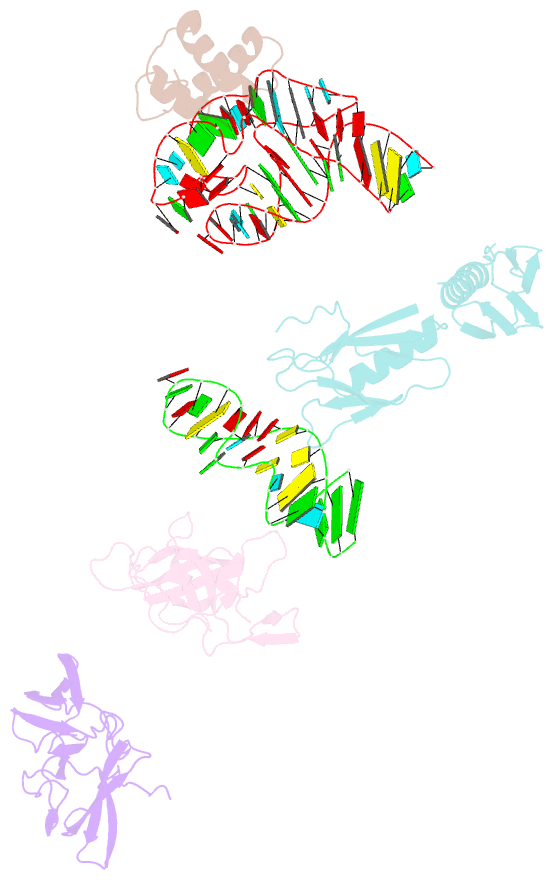
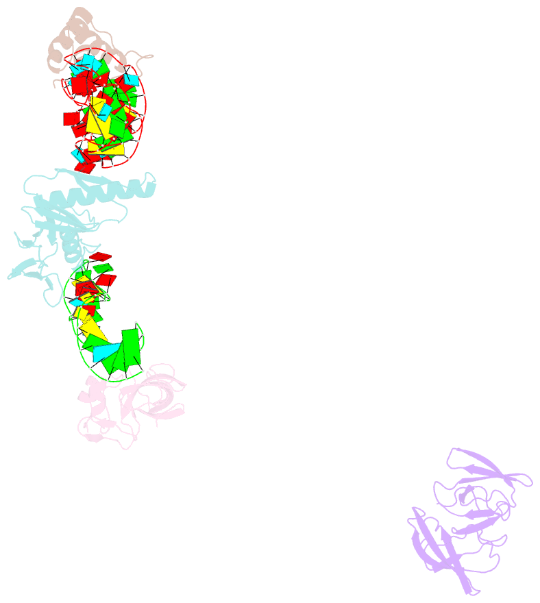
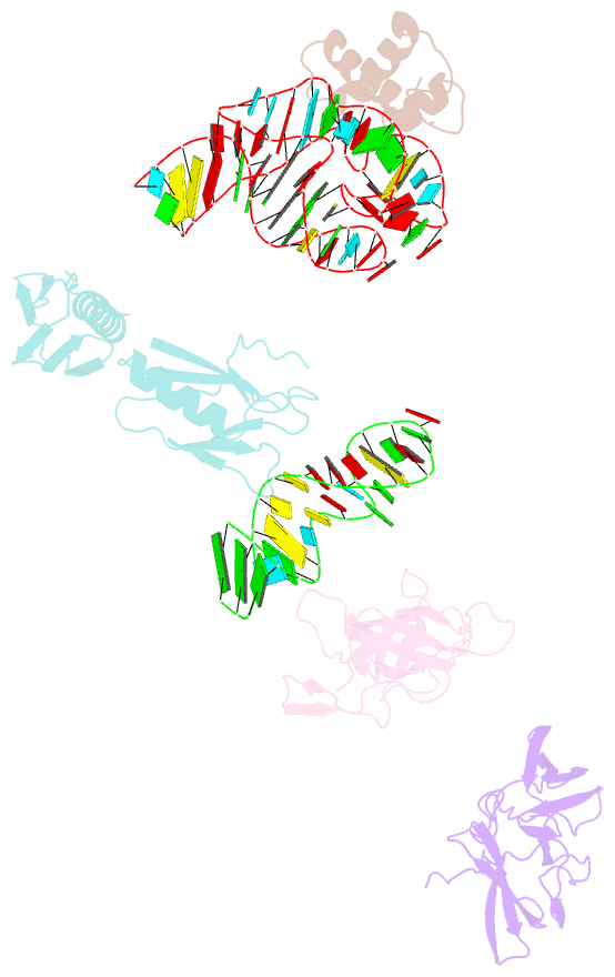
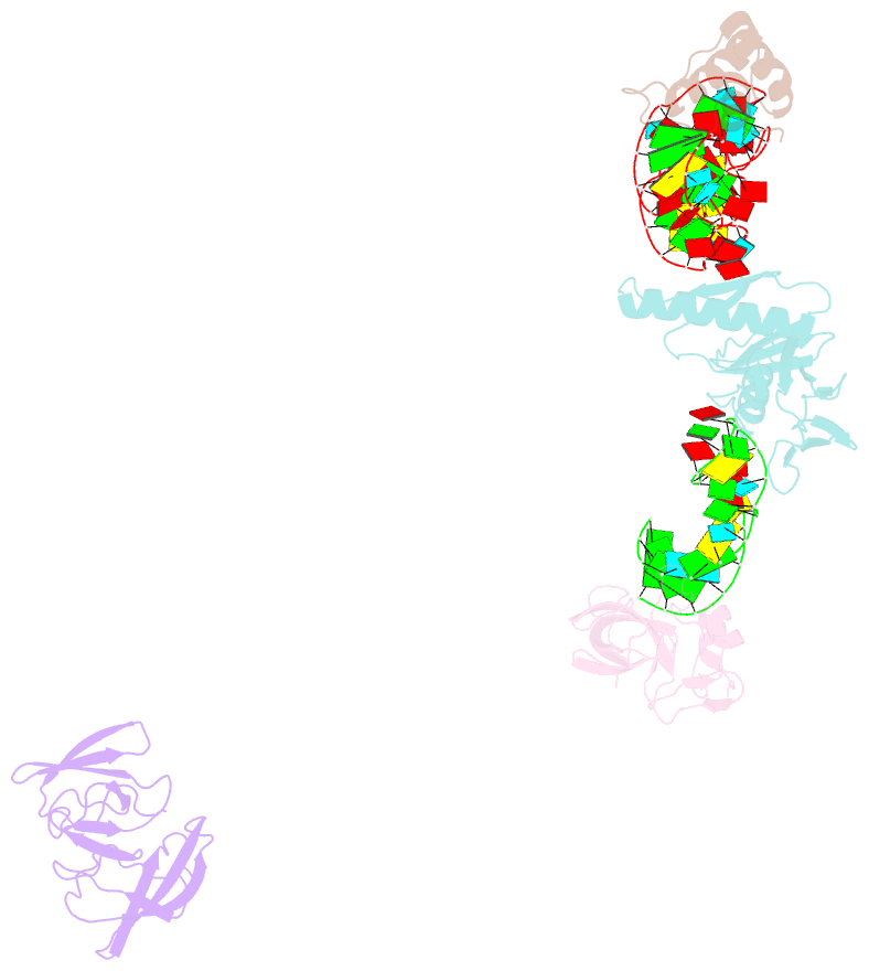
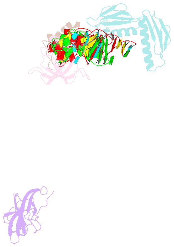
- Identification of known protein and RNA structures in a 5 a map of the large ribosomal subunit from haloarcula marismortui
- Reference
- Ban N, Nissen P, Hansen J, Capel M, Moore PB, Steitz TA (1999): "Placement of protein and RNA structures into a 5 A-resolution map of the 50S ribosomal subunit." Nature, 400, 841-847. doi: 10.1038/23641.
- Abstract
- We have calculated at 5.0 A resolution an electron-density map of the large 50S ribosomal subunit from the bacterium Haloarcula marismortui by using phases derived from four heavy-atom derivatives, intercrystal density averaging and density-modification procedures. More than 300 base pairs of A-form RNA duplex have been fitted into this map, as have regions of non-A-form duplex, single-stranded segments and tetraloops. The long rods of RNA crisscrossing the subunit arise from the stacking of short, separate double helices, not all of which are A-form, and in many places proteins crosslink two or more of these rods. The polypeptide exit channel was marked by tungsten cluster compounds bound in one heavy-atom-derivatized crystal. We have determined the structure of the translation-factor-binding centre by fitting the crystal structures of the ribosomal proteins L6, L11 and L14, the sarcin-ricin loop RNA, and the RNA sequence that binds L11 into the electron density. We can position either elongation factor G or elongation factor Tu complexed with an aminoacylated transfer RNA and GTP onto the factor-binding centre in a manner that is consistent with results from biochemical and electron microscopy studies.