Summary information and primary citation
- PDB-id
- 1c0w; SNAP-derived features in text and JSON formats;
DNAproDB
- Class
- gene regulation-DNA
- Method
- X-ray (3.2 Å)
- Summary
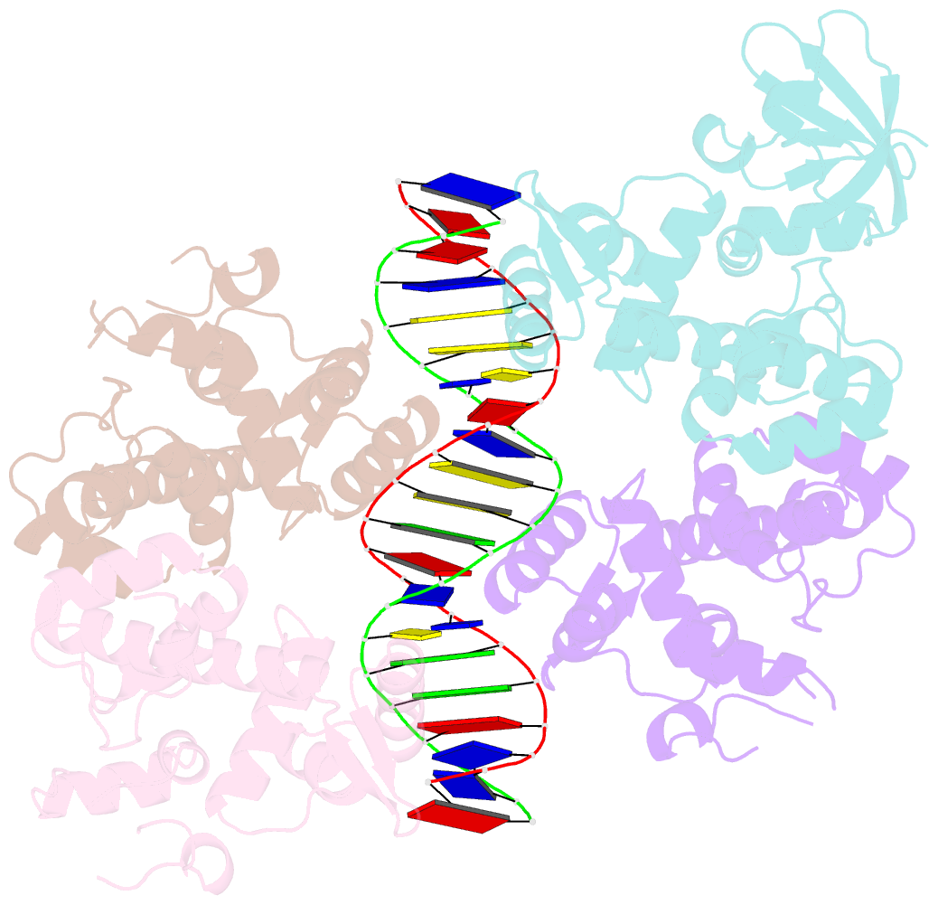
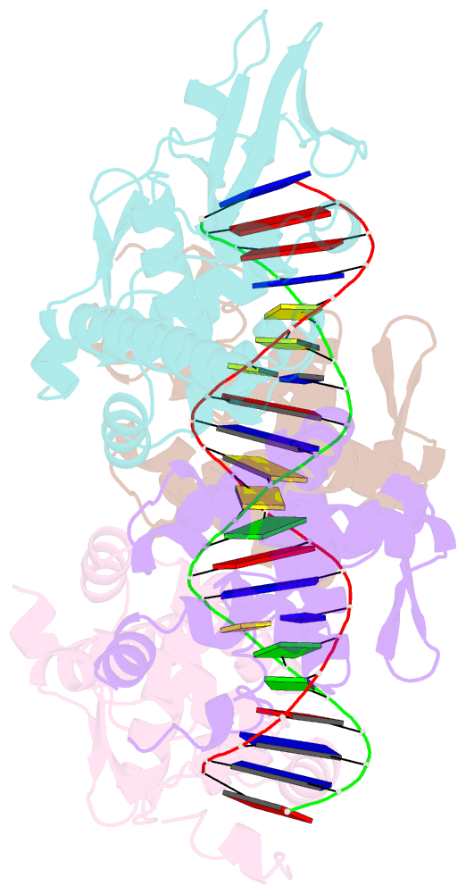
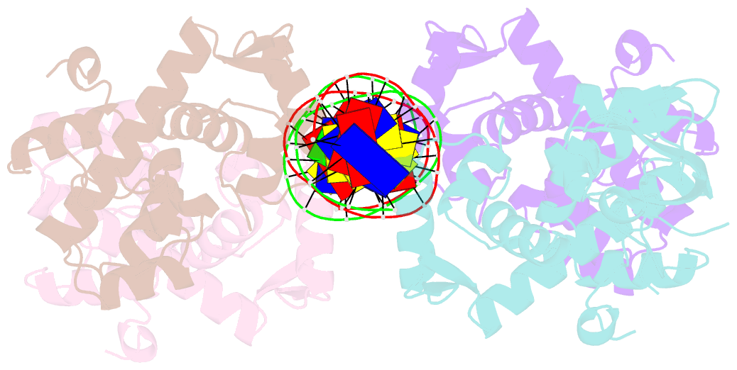
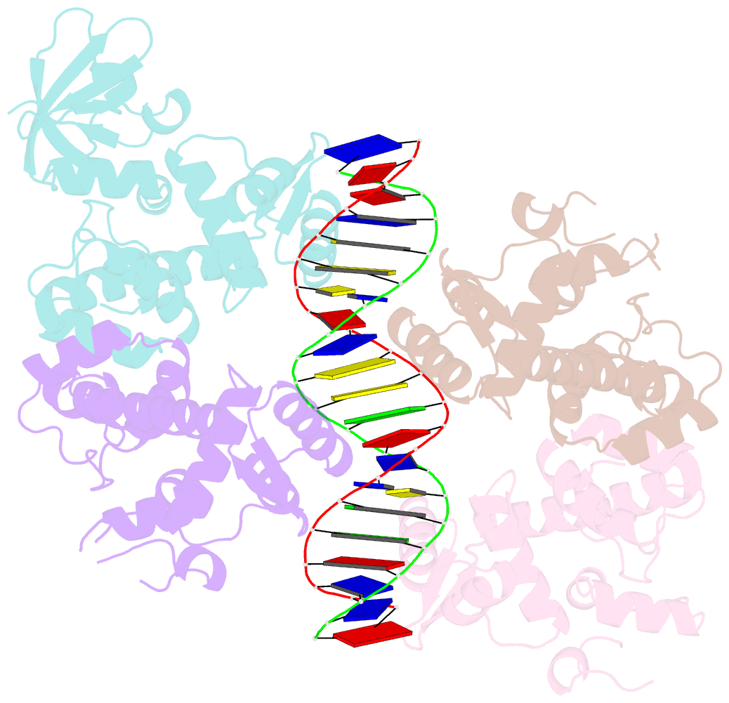
- Crystal structure of the cobalt-activated diphtheria toxin repressor-DNA complex reveals a metal binding sh-like domain
- Reference
- Pohl E, Holmes RK, Hol WG (1999): "Crystal structure of a cobalt-activated diphtheria toxin repressor-DNA complex reveals a metal-binding SH3-like domain." J.Mol.Biol., 292, 653-667. doi: 10.1006/jmbi.1999.3073.
- Abstract
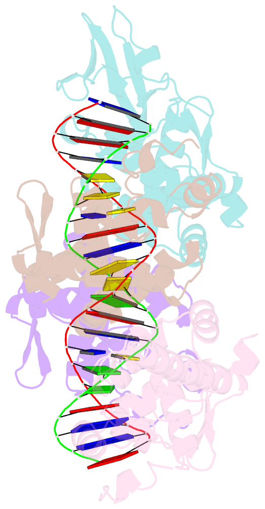
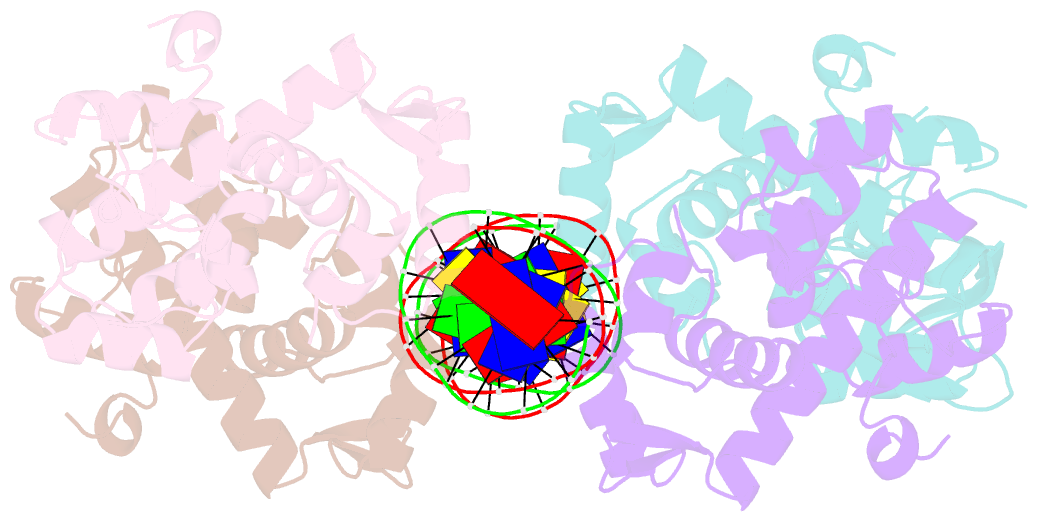
- The diphtheria toxin repressor (DtxR) is the prototype of a family of iron-dependent regulator (IdeR) proteins, which are activated by divalent iron and bind DNA to prevent the transcription of downstream genes. In Corynebacterium diphtheriae, DtxR regulates not only the expression of diphtheria toxin encoded by a corynebacteriophage, but also of components of the siderophore-mediated iron-transport system. Here we report the crystal structure of wild-type DtxR, a 226 residue three-domain dimeric protein, activated by cobalt and bound to a 21 bp DNA duplex based on the consensus operator sequence. Two DtxR dimers surround the DNA duplex which is distorted compared to canonical B -DNA. The SH3-like third domain interacts with the metal at site 1 via the side-chains of Glu170 and Gln173, revealing for the first time a metal-binding function for this class of domains. The SH3-like domain is also in contact with the DNA-binding first domain and with the second, or dimerization, domain. The DNA-binding helices in the first domain are shifted by 3 to 5 A when compared to the apo-repressor, and fit into the major groove of the duplex bound. These shifts are due to a hinge-binding motion of the DNA-binding domain with respect to the dimerization domains of DtxR. The third domain might play a role in regulating this hinge motion.