Summary information and primary citation
- PDB-id
- 1cjg; SNAP-derived features in text and JSON formats;
DNAproDB
- Class
- transcription-DNA
- Method
- NMR
- Summary
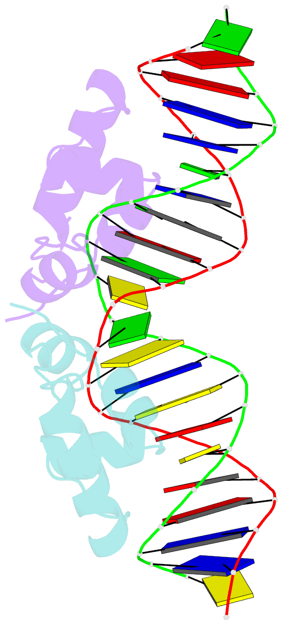
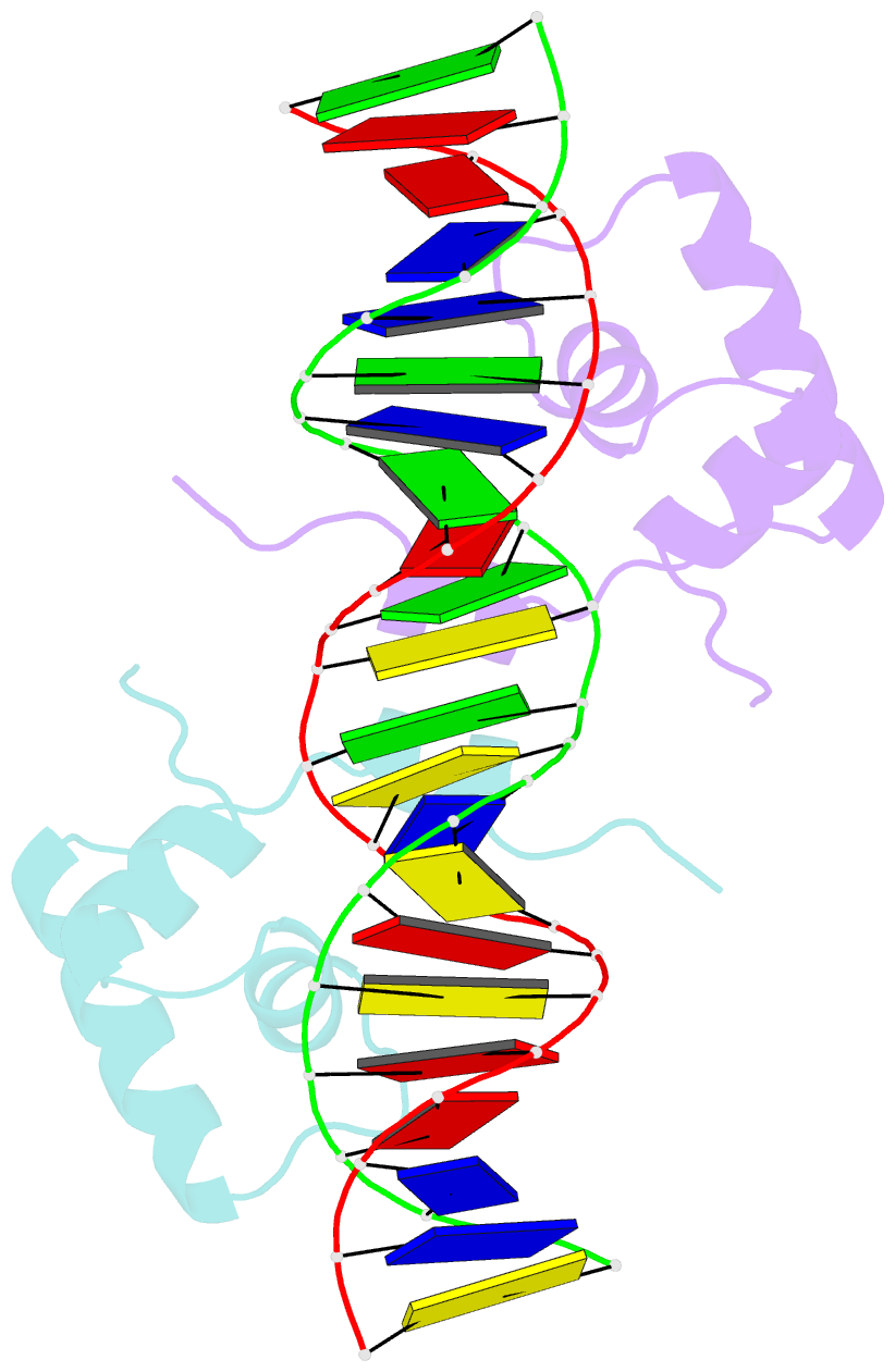
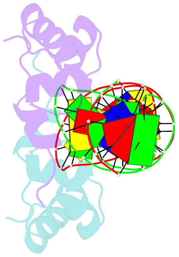
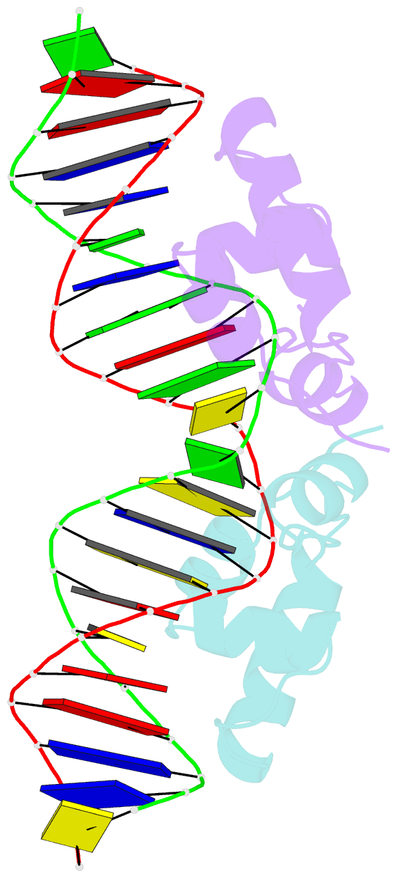
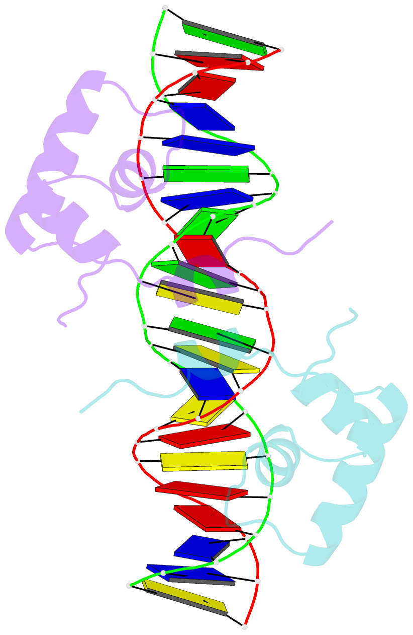
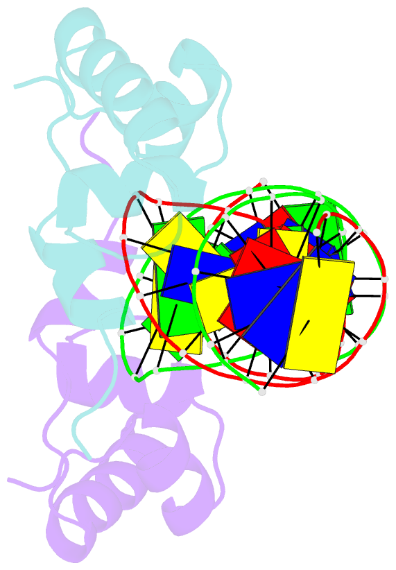
- NMR structure of lac repressor hp62-DNA complex
- Reference
- Spronk CA, Bonvin AM, Radha PK, Melacini G, Boelens R, Kaptein R (1999): "The solution structure of Lac repressor headpiece 62 complexed to a symmetrical lac operator." Structure Fold.Des., 7, 1483-1492. doi: 10.1016/S0969-2126(00)88339-2.
- Abstract
- Background: Lactose repressor protein (Lac) controls the expression of the lactose metabolic genes in Escherichia coli by binding to an operator sequence in the promoter of the lac operon. Binding of inducer molecules to the Lac core domain induces changes in tertiary structure that are propagated to the DNA-binding domain through the connecting hinge region, thereby reducing the affinity for the operator. Protein-protein and protein-DNA interactions involving the hinge region play a crucial role in the allosteric changes occurring upon induction, but have not, as yet, been analyzed in atomic detail.
Results: We have used nuclear magnetic resonance (NMR) spectroscopy and restrained molecular dynamics (rMD) to determine the structure of the Lac repressor DNA-binding domain (headpeice 62; HP62) in complex with a symmetrized lac operator. Analysis of the structures reveals specific interactions between Lac repressor and DNA that were not found in previously investigated Lac repressor-DNA complexes. Important differences with the previously reported structures of the HP56-DNA complex were found in the loop following the helix-turn-helix (HTH) motif. The protein-protein and protein-DNA interactions involving the hinge region and the deformations in the DNA structure could be delineated in atomic detail. The structures were also used for comparison with the available crystallographic data on the Lac and Pur repressor-DNA complexes.
Conclusions: The structures of the HP62-DNA complex provide the basis for a better understanding of the specific recognition in the Lac repressor-operator complex. In addition, the structural features of the hinge region provide detailed insight into the protein-protein and protein-DNA interactions responsible for the high affinity of the repressor for operator DNA.