Summary information and primary citation
- PDB-id
- 1cma; SNAP-derived features in text and JSON formats;
DNAproDB
- Class
- transcription-DNA
- Method
- X-ray (2.8 Å)
- Summary
- Met repressor-DNA complex + s-adenosyl-methionine
- Reference
- Somers WS, Phillips SE (1992): "Crystal structure of the met repressor-operator complex at 2.8 A resolution reveals DNA recognition by beta-strands." Nature, 359, 387-393. doi: 10.1038/359387a0.
- Abstract
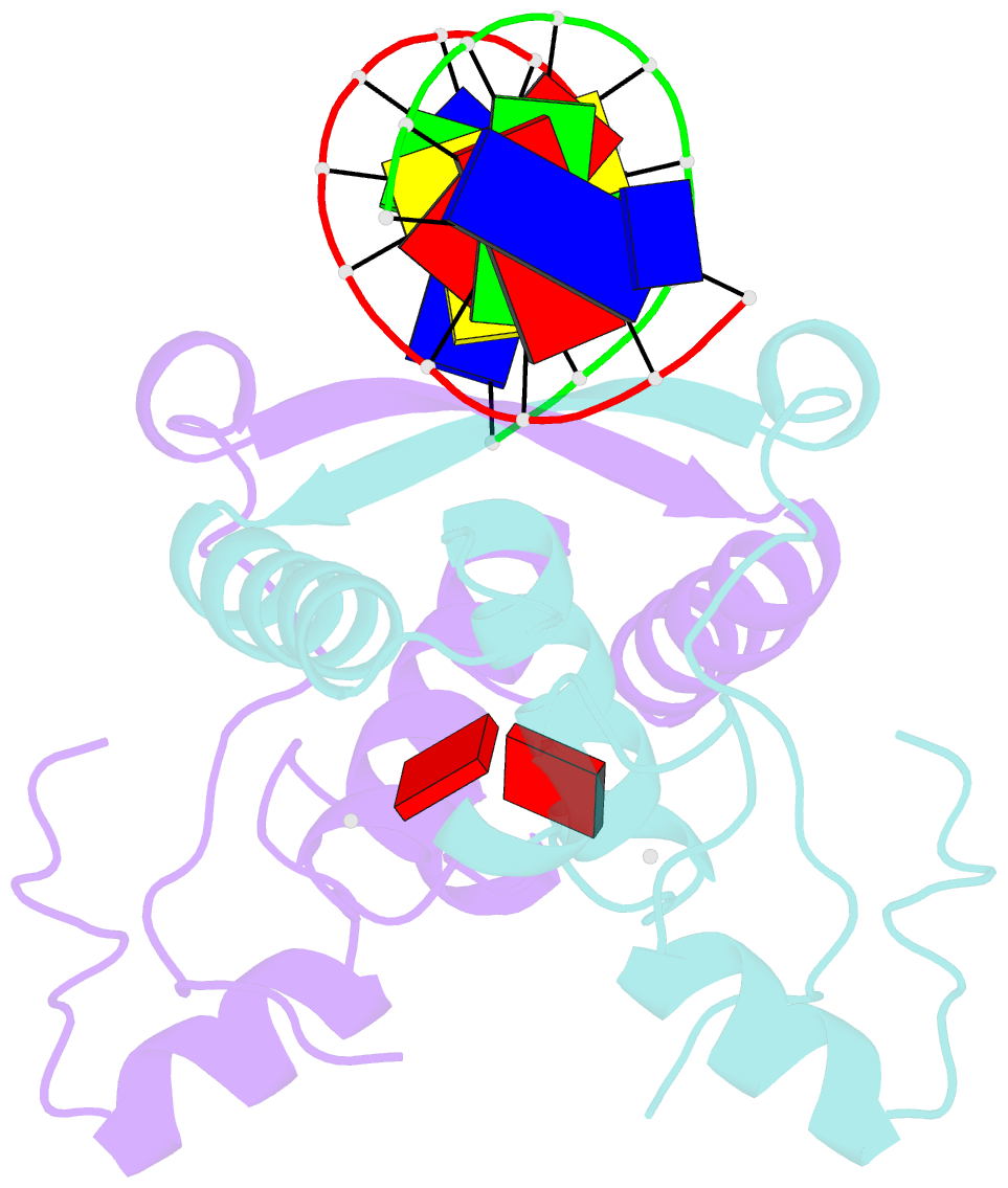
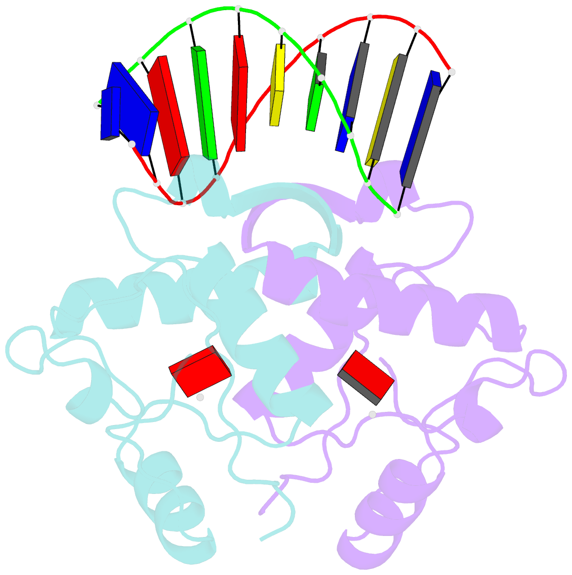
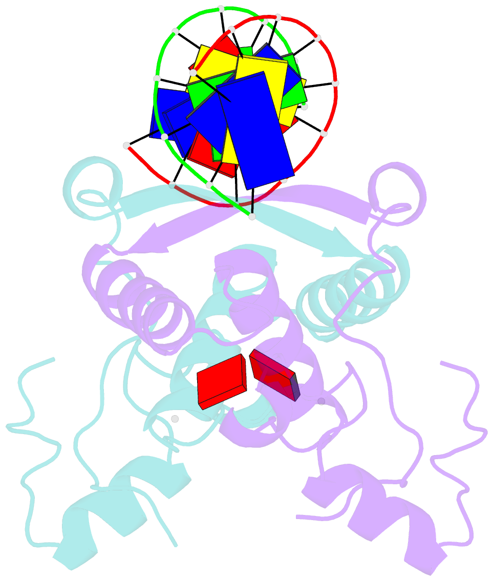
- The crystal structure of the met repressor-operator complex shows two dimeric repressor molecules bound to adjacent sites 8 base pairs apart on an 18-base-pair DNA fragment. Sequence specificity is achieved by insertion of double-stranded antiparallel protein beta-ribbons into the major groove of B-form DNA, with direct hydrogen-bonding between amino-acid side chains and the base pairs. The repressor also recognizes sequence-dependent distortion or flexibility of the operator phosphate backbone, conferring specificity even for inaccessible base pairs.