Summary information and primary citation
- PDB-id
- 1cvj; SNAP-derived features in text and JSON formats;
DNAproDB
- Class
- gene regulation-RNA
- Method
- X-ray (2.6 Å)
- Summary
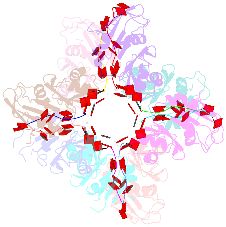
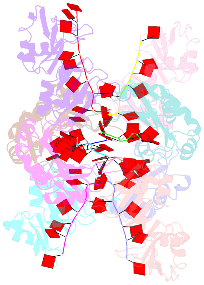
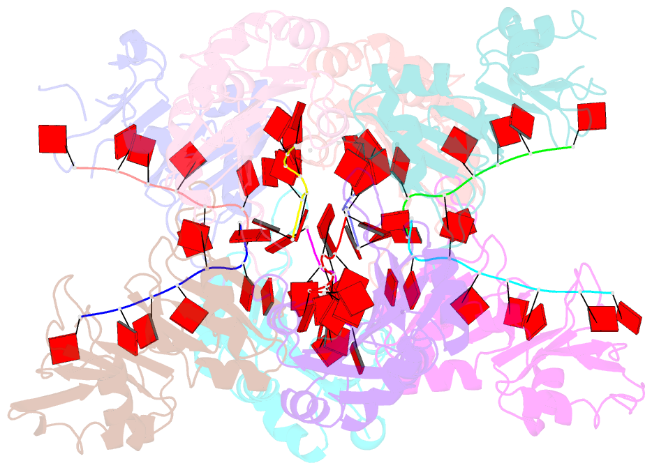
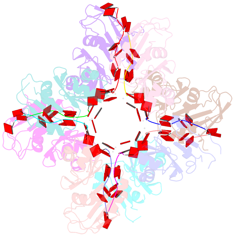
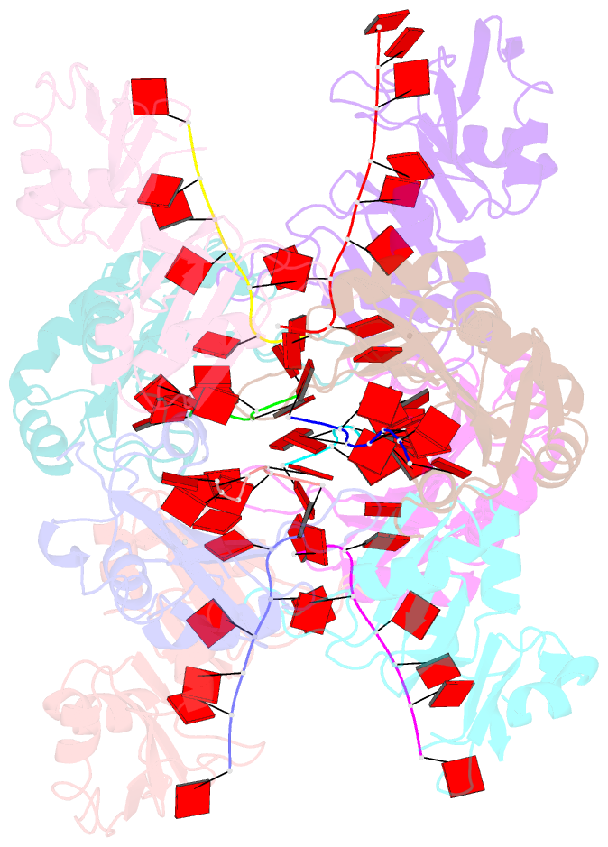
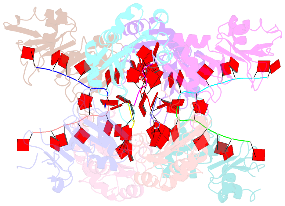
- X-ray crystal structure of the poly(a)-binding protein in complex with polyadenylate RNA
- Reference
- Deo RC, Bonanno JB, Sonenberg N, Burley SK (1999): "Recognition of polyadenylate RNA by the poly(A)-binding protein." Cell(Cambridge,Mass.), 98, 835-845. doi: 10.1016/S0092-8674(00)81517-2.
- Abstract
- The cocrystal structure of human poly(A)-binding protein (PABP) has been determined at 2.6 A resolution. PABP recognizes the 3' mRNA poly(A) tail and plays critical roles in eukaryotic translation initiation and mRNA stabilization/degradation. The minimal PABP used in this study consists of the N-terminal two RRM-type RNA-binding domains connected by a short linker (RRM1/2). These two RRMs form a continuous RNA-binding trough, lined by an antiparallel beta sheet backed by four alpha helices. The polyadenylate RNA adopts an extended conformation running the length of the molecular trough. Adenine recognition is primarily mediated by contacts with conserved residues found in the RNP motifs of the two RRMs. The convex dorsum of RRM1/2 displays a phylogenetically conserved hydrophobic/acidic portion, which may interact with translation initiation factors and regulatory proteins.