Summary information and primary citation
- PDB-id
- 1d0e; SNAP-derived features in text and JSON formats;
DNAproDB
- Class
- transferase-DNA
- Method
- X-ray (3.0 Å)
- Summary
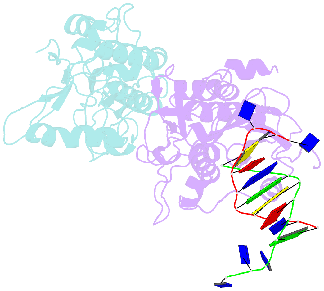
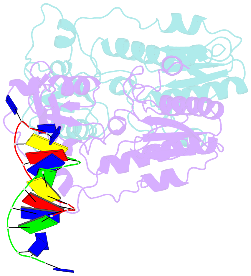
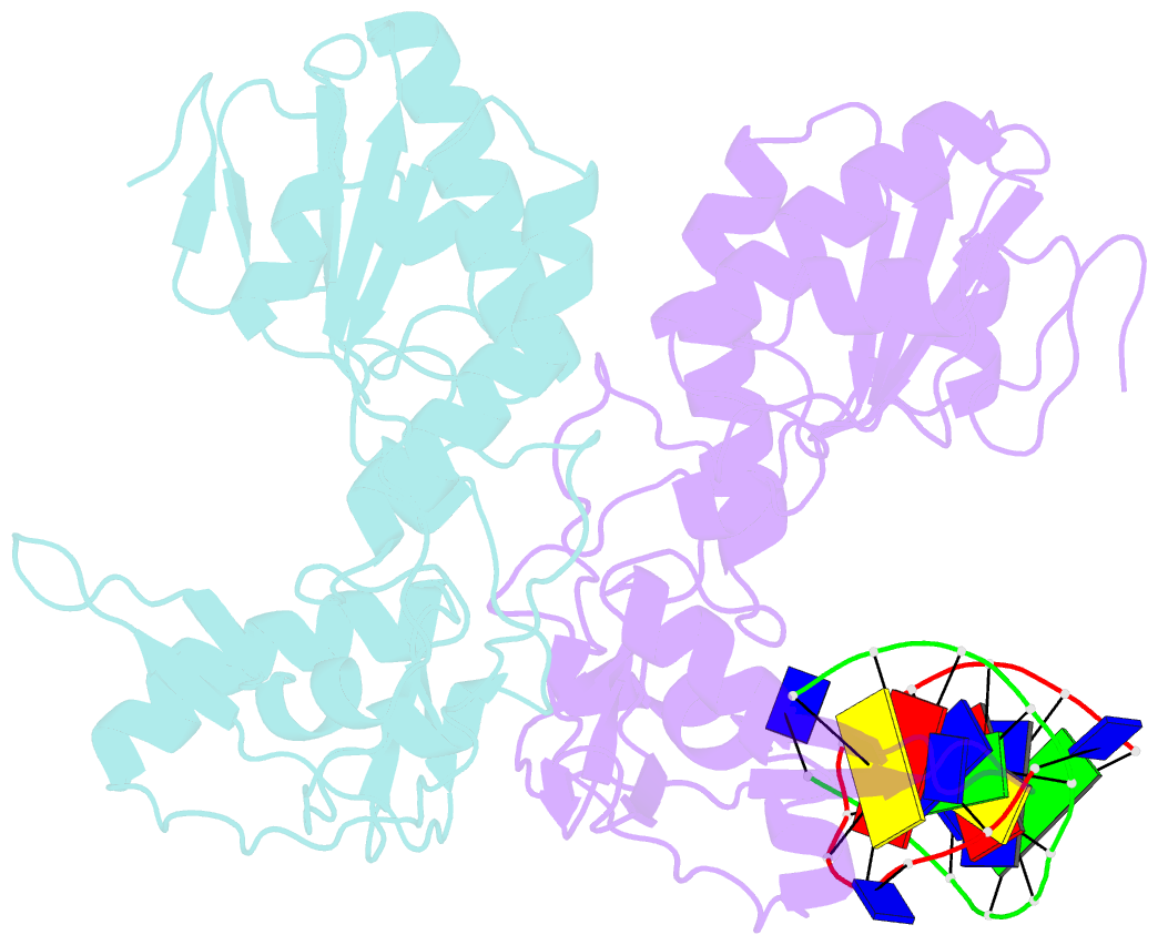
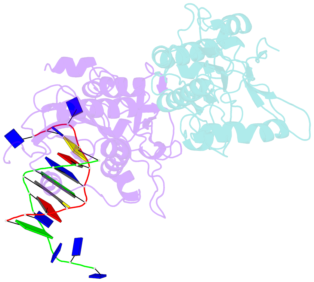
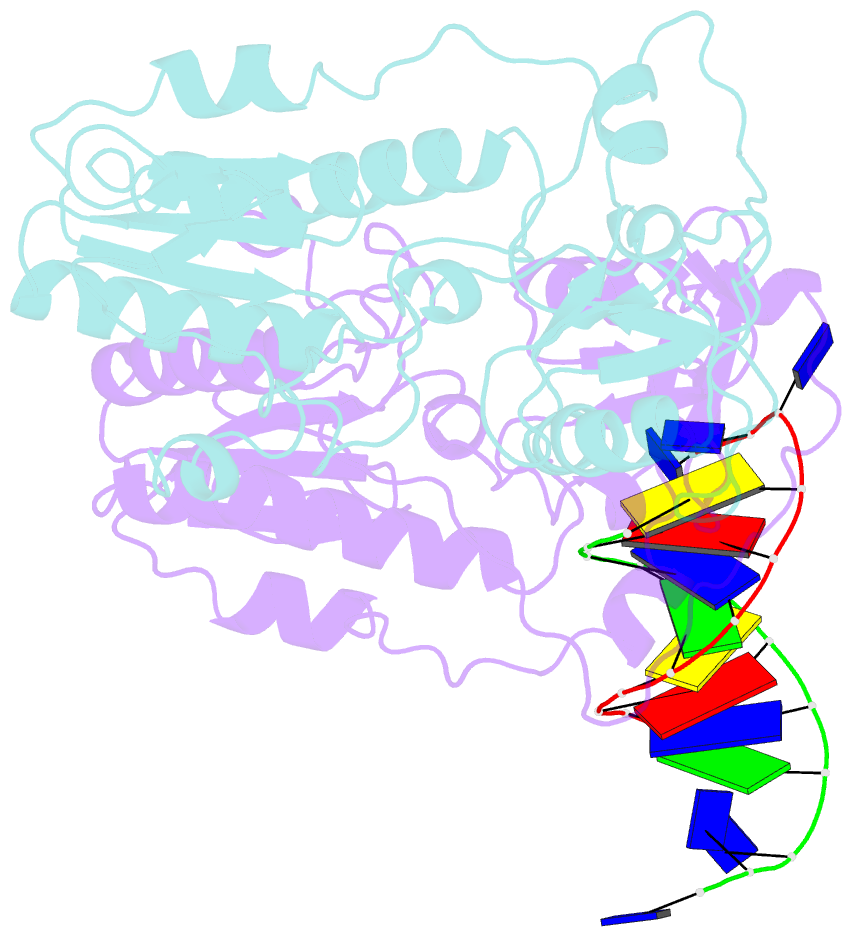
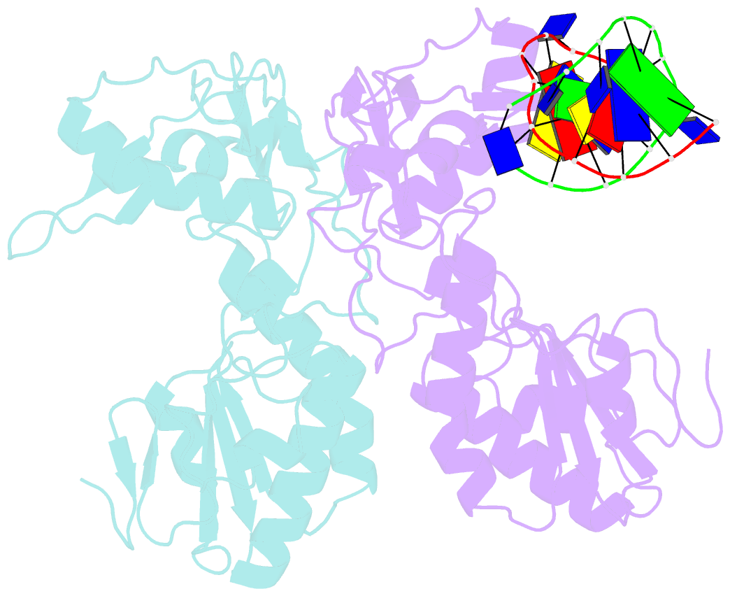
- Crystal structures of the n-terminal fragment from moloney murine leukemia virus reverse transcriptase complexed with nucleic acid: functional implications for template-primer binding to the fingers domain
- Reference
- Najmudin S, Cote ML, Sun D, Yohannan S, Montano SP, Gu J, Georgiadis MM (2000): "Crystal structures of an N-terminal fragment from Moloney murine leukemia virus reverse transcriptase complexed with nucleic acid: functional implications for template-primer binding to the fingers domain." J.Mol.Biol., 296, 613-632. doi: 10.1006/jmbi.1999.3477.
- Abstract
- Reverse transcriptase (RT) serves as the replicative polymerase for retroviruses by using RNA and DNA-directed DNA polymerase activities coupled with a ribonuclease H activity to synthesize a double-stranded DNA copy of the single-stranded RNA genome. In an effort to obtain detailed structural information about nucleic acid interactions with reverse transcriptase, we have determined crystal structures at 2.3 A resolution of an N-terminal fragment from Moloney murine leukemia virus reverse transcriptase complexed to blunt-ended DNA in three distinct lattices. This fragment includes the fingers and palm domains from Moloney murine leukemia virus reverse transcriptase. We have also determined the crystal structure at 3.0 A resolution of the fragment complexed to DNA with a single-stranded template overhang resembling a template-primer substrate. Protein-DNA interactions, which are nearly identical in each of the three lattices, involve four conserved residues in the fingers domain, Asp114, Arg116, Asn119 and Gly191. DNA atoms involved in the interactions include the 3'-OH group from the primer strand and minor groove base atoms and sugar atoms from the n-2 and n-3 positions of the template strand, where n is the template base that would pair with an incoming nucleotide. The single-stranded template overhang adopts two different conformations in the asymmetric unit interacting with residues in the beta4-beta5 loop (beta3-beta4 in HIV-1 RT). Our fragment-DNA complexes are distinct from previously reported complexes of DNA bound to HIV-1 RT but related in the types of interactions formed between protein and DNA. In addition, the DNA in all of these complexes is bound in the same cleft of the enzyme. Through site-directed mutagenesis, we have substituted residues that are involved in binding DNA in our crystal structures and have characterized the resulting enzymes. We now propose that nucleic acid binding to the fingers domain may play a role in translocation of nucleic acid during processive DNA synthesis and suggest that our complex may represent an intermediate in this process.