Summary information and primary citation
- PDB-id
- 1d6k; SNAP-derived features in text and JSON formats;
DNAproDB
- Class
- ribosome
- Method
- NMR
- Summary
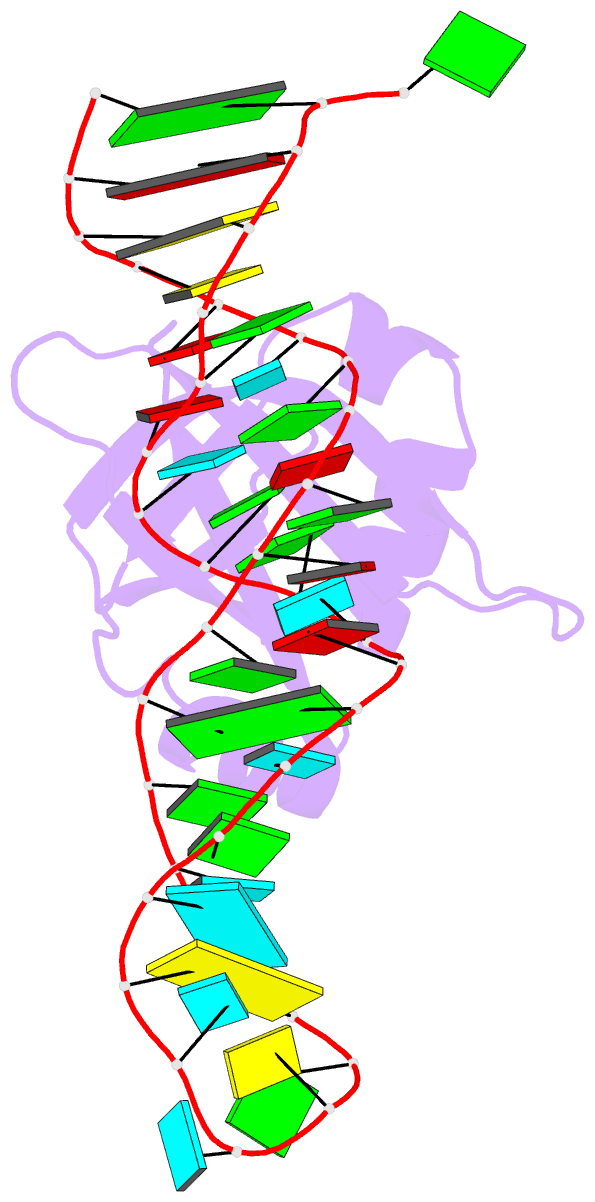
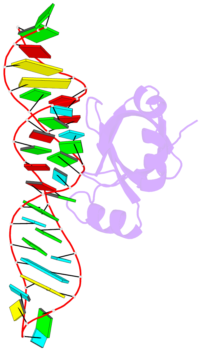
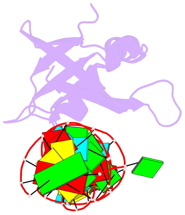
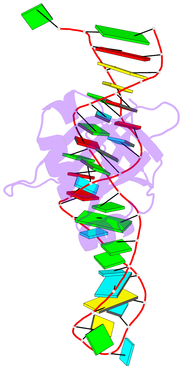
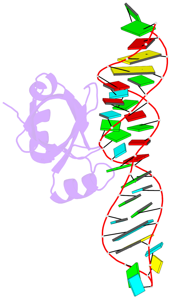
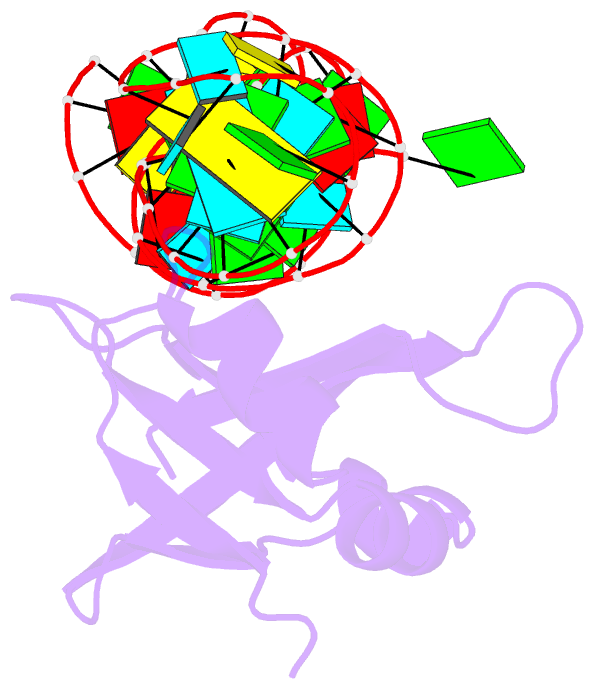
- NMR solution structure of the 5s rrna e-loop-l25 complex
- Reference
- Stoldt M, Wohnert J, Ohlenschlager O, Gorlach M, Brown LR (1999): "The NMR structure of the 5S rRNA E-domain-protein L25 complex shows preformed and induced recognition." EMBO J., 18, 6508-6521. doi: 10.1093/emboj/18.22.6508.
- Abstract
- The structure of the complex between ribosomal protein L25 and a 37 nucleotide RNA molecule, which contains the E-loop and helix IV regions of the E-domain of Escherichia coli 5S rRNA, has been determined to an overall r.m.s. displacement of 1.08 A (backbone heavy atoms) by heteronuclear NMR spectroscopy (Protein Databank code 1d6k). The interacting molecular surfaces are bipartite for both the RNA and the protein. One side of the six-stranded beta-barrel of L25 recognizes the minor groove of the E-loop with very little change in the conformations of either the protein or the RNA and with the RNA-protein interactions occurring mainly along one strand of the E-loop duplex. This minor groove recognition module includes two parallel beta-strands of L25, a hitherto unknown RNA binding topology. Binding of the RNA also induces conversion of a flexible loop to an alpha-helix in L25, the N-terminal tip of which interacts with the widened major groove at the E-loop/helix IV junction of the RNA. The structure of the complex reveals that the E-domain RNA serves as a preformed docking partner, while the L25 protein has one preformed and one induced recognition module.