Summary information and primary citation
- PDB-id
- 1d9f; SNAP-derived features in text and JSON formats;
DNAproDB
- Class
- transferase-DNA, RNA
- Method
- X-ray (3.0 Å)
- Summary
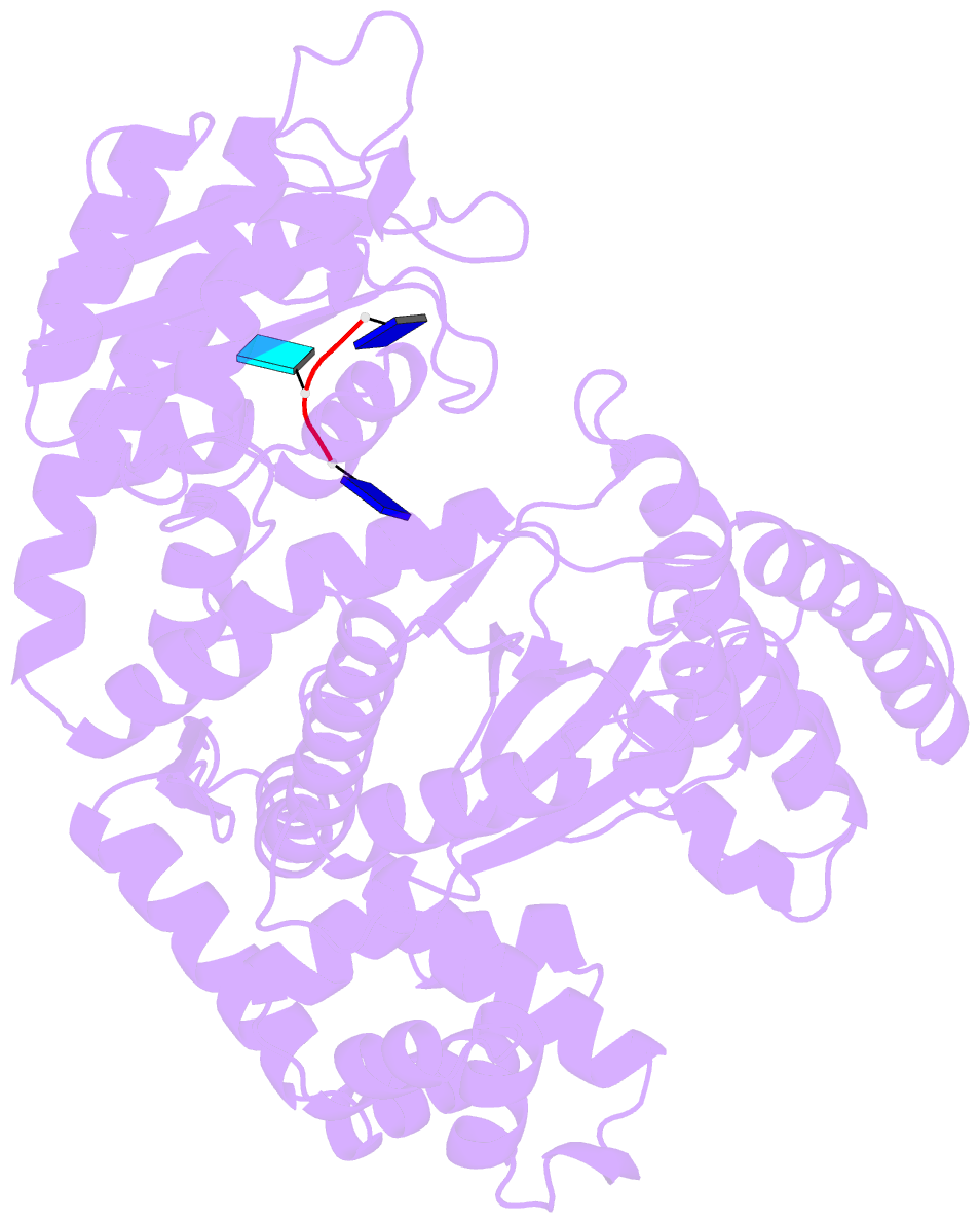
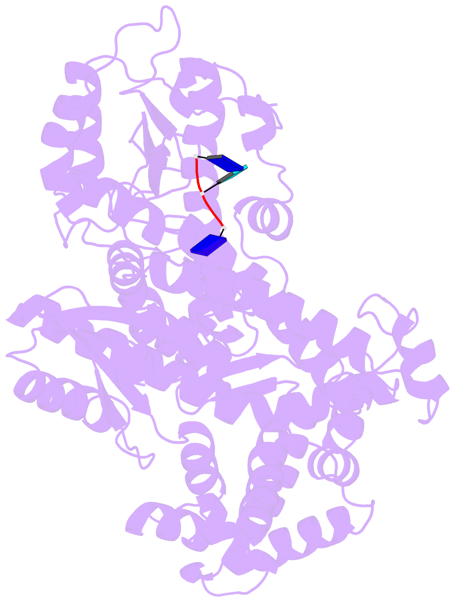
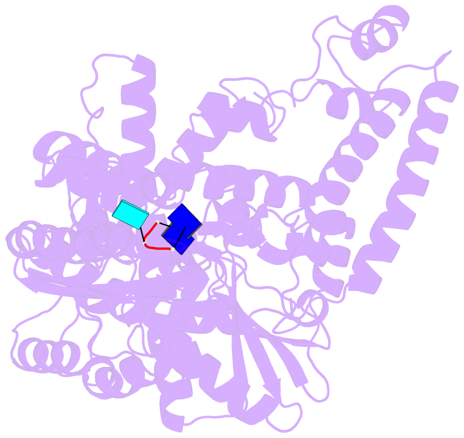
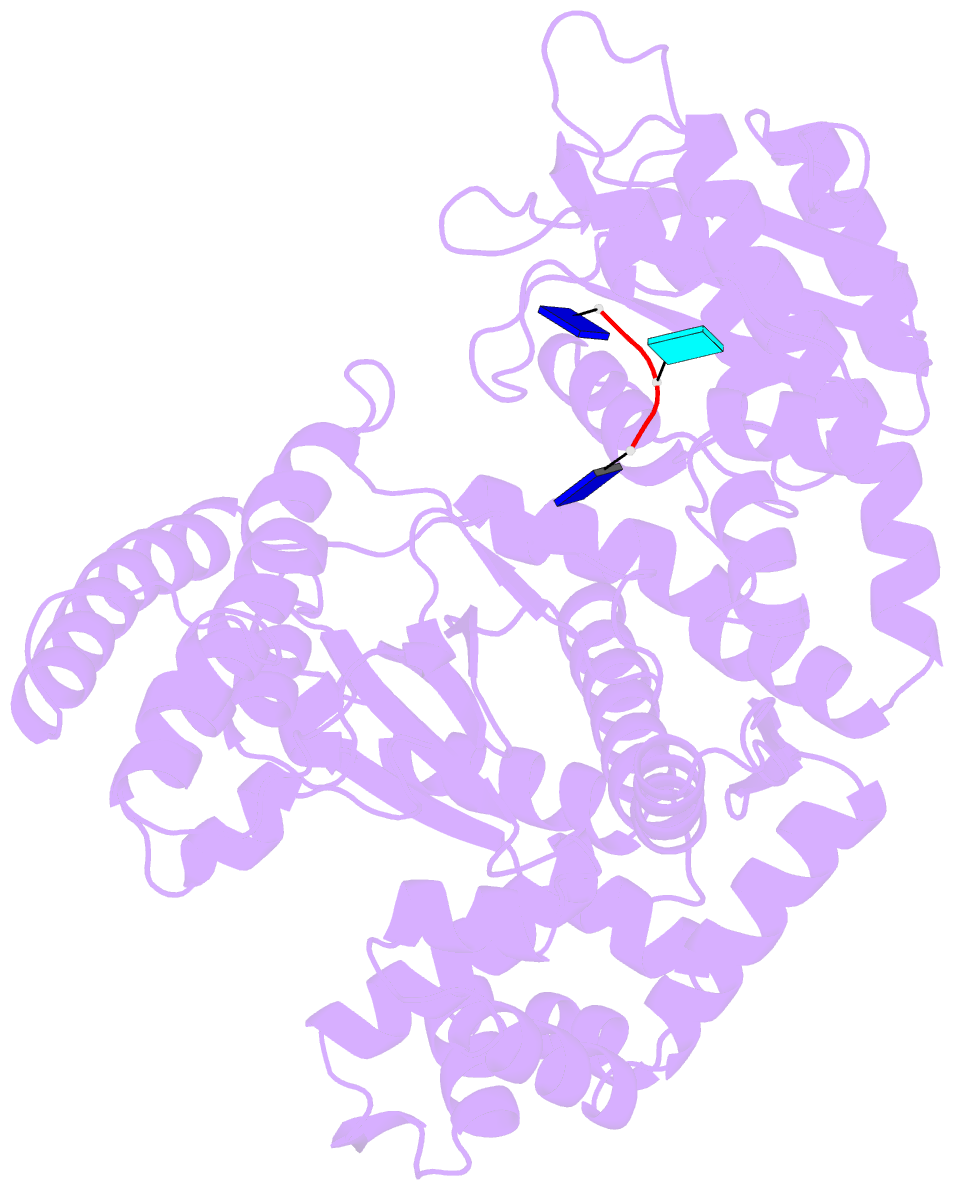
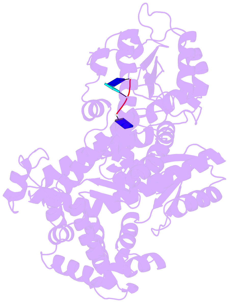
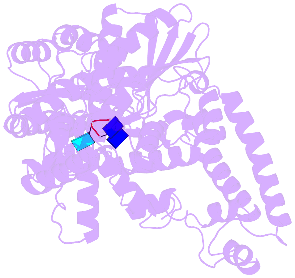
- Crystal structure of the complex of DNA polymerase i klenow fragment with DNA tetramer carrying 2'-o-(3-aminopropyl)-RNA modification 5'-d(tt)-ap(u)-d(t)-3'
- Reference
- Teplova M, Wallace ST, Tereshko V, Minasov G, Symons AM, Cook PD, Manoharan M, Egli M (1999): "Structural origins of the exonuclease resistance of a zwitterionic RNA." Proc.Natl.Acad.Sci.USA, 96, 14240-14245. doi: 10.1073/pnas.96.25.14240.
- Abstract
- Nuclease resistance and RNA affinity are key criteria in the search for optimal antisense nucleic acid modifications, but the origins of the various levels of resistance to nuclease degradation conferred by chemical modification of DNA and RNA are currently not understood. The 2'-O-aminopropyl (AP)-RNA modification displays the highest nuclease resistance among all phosphodiester-based analogues and its RNA binding affinity surpasses that of phosphorothioate DNA by 1 degrees C per modified residue. We found that oligodeoxynucleotides containing AP-RNA residues at their 3' ends competitively inhibit the degradation of single-stranded DNA by the Escherichia coli Klenow fragment (KF) 3'-5' exonuclease and snake venom phosphodiesterase. To shed light on the origins of nuclease resistance brought about by the AP modification, we determined the crystal structure of an A-form DNA duplex with AP-RNA modifications at 1.6-A resolution. In addition, the crystal structures of complexes between short DNA fragments carrying AP-RNA modifications and wild-type KF were determined at resolutions between 2.2 and 3.0 A and compared with the structure of the complex between oligo(dT) and the D355A/E357A KF mutant. The structural models suggest that interference of the positively charged 2'-O-substituent with the metal ion binding site B of the exonuclease allows AP-RNA to effectively slow down degradation.