Summary information and primary citation
- PDB-id
- 1ddn; SNAP-derived features in text and JSON formats;
DNAproDB
- Class
- gene regulation-DNA
- Method
- X-ray (3.0 Å)
- Summary
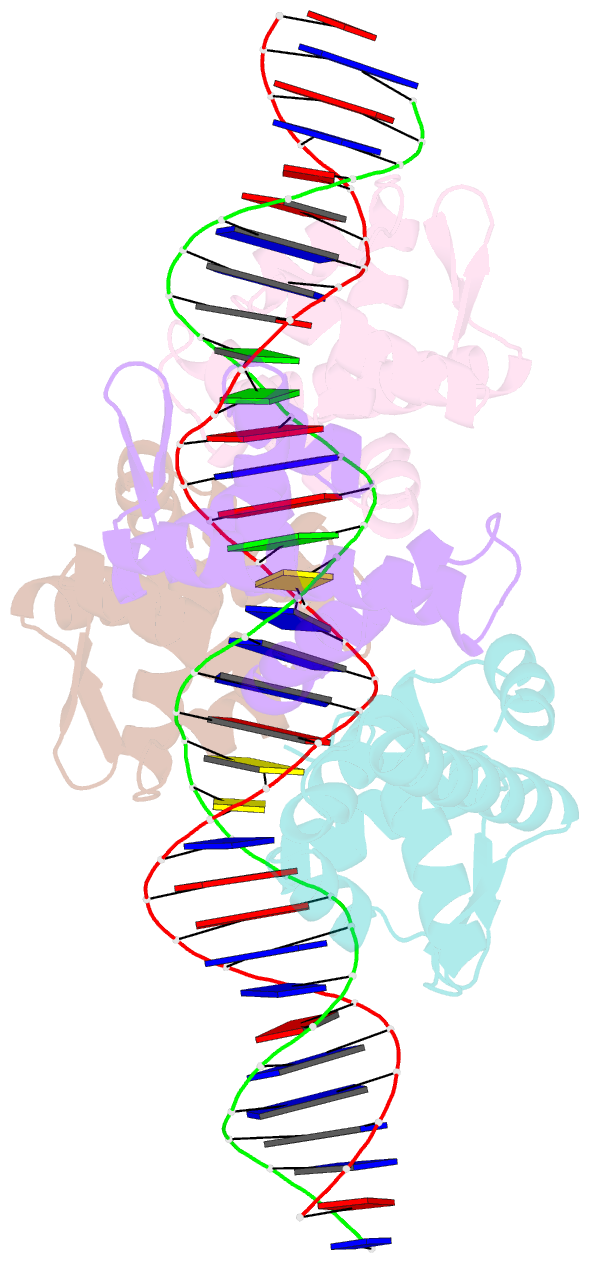
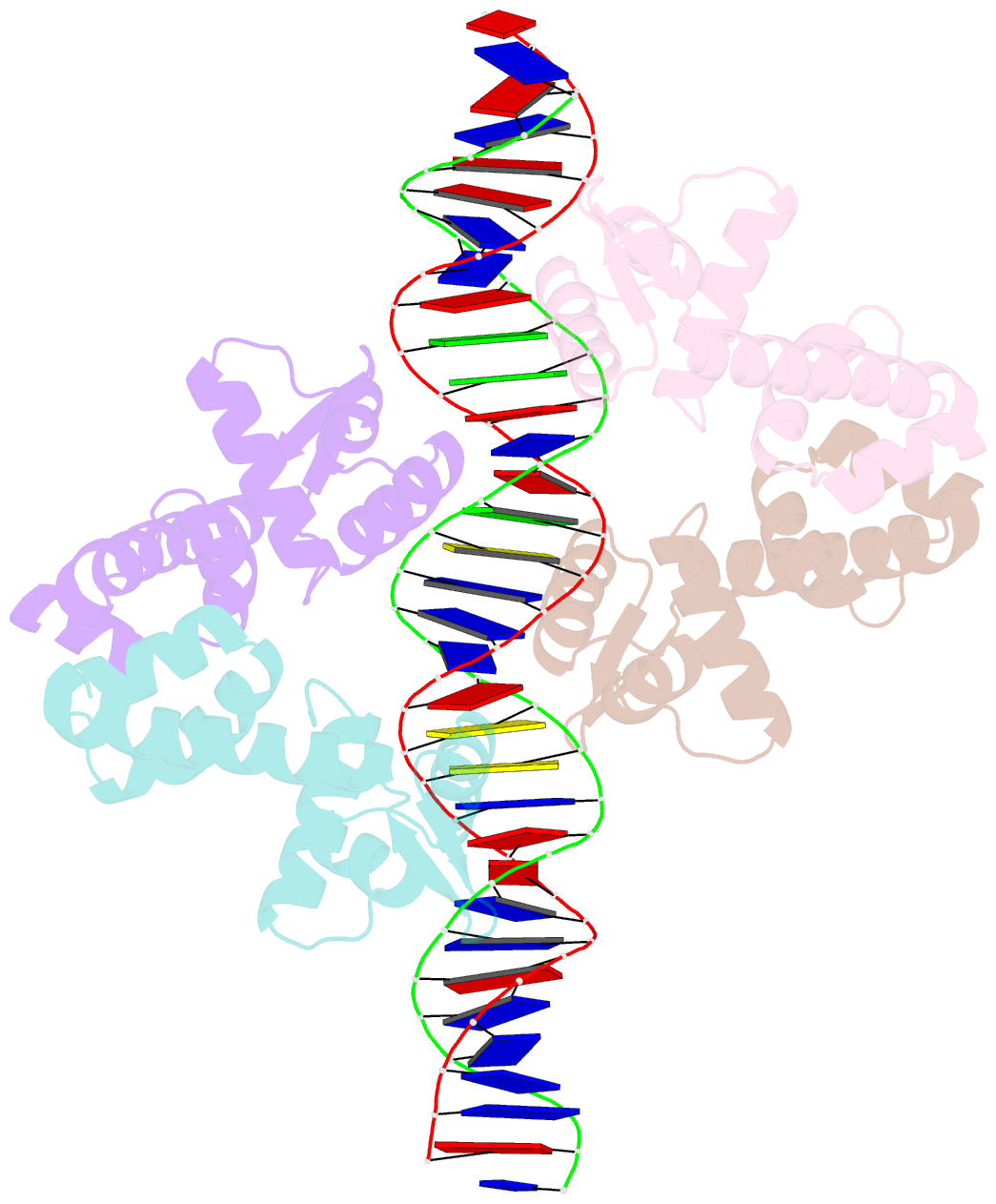
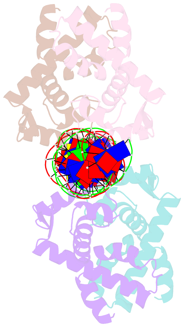
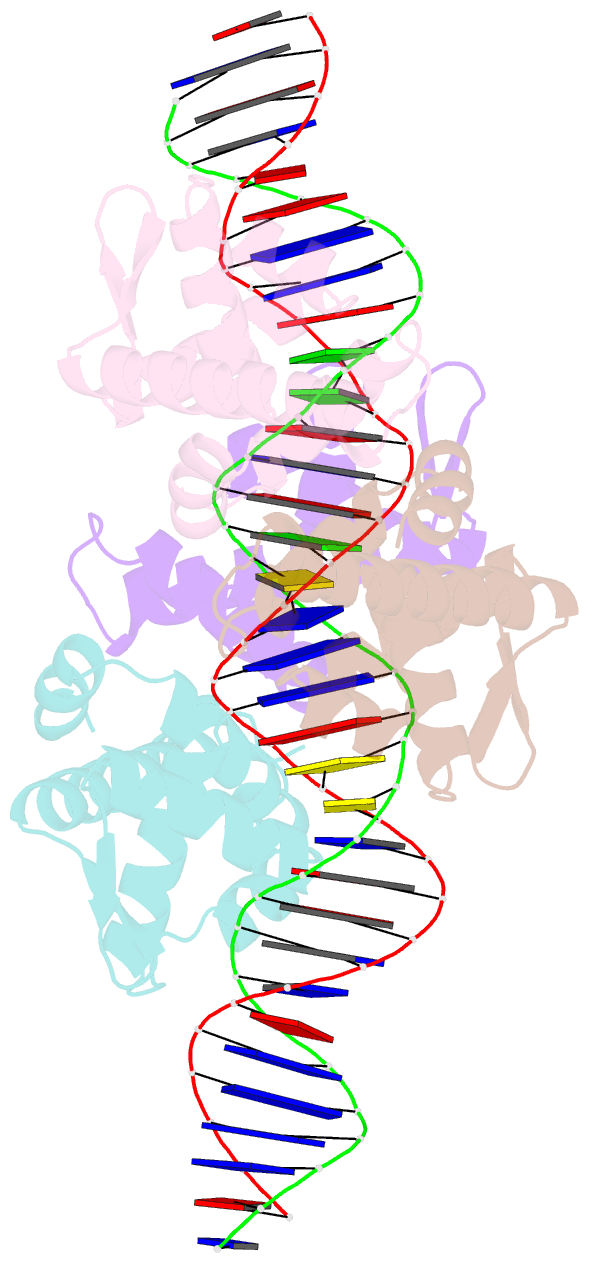
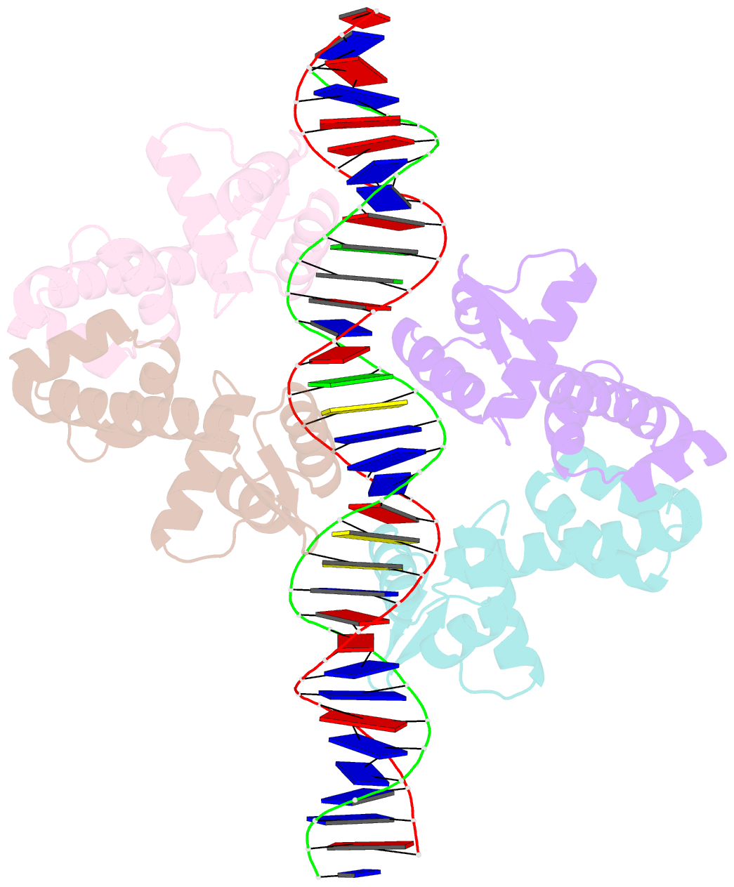
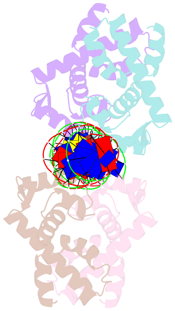
- Diphtheria tox repressor (c102d mutant)-tox DNA operator complex
- Reference
- White A, Ding X, vanderSpek JC, Murphy JR, Ringe D (1998): "Structure of the metal-ion-activated diphtheria toxin repressor/tox operator complex." Nature, 394, 502-506. doi: 10.1038/28893.
- Abstract
- The virulent phenotype of the pathogenic bacterium Corynebacterium diphtheriae is conferred by diphtheria toxin, whose expression is an adaptive response to low concentrations of iron. The expression of the toxin gene (tox) is regulated by the repressor DtxR, which is activated by transition metal ions. X-ray crystal structures of DtxR with and without (apo-form) its coordinated transition metal ion have established the general architecture of the repressor, identified the location of the metal-binding sites, and revealed a metal-ion-triggered subunit-subunit 'caliper-like' conformational change. Here we report the three-dimensional crystal structure of the complex between a biologically active Ni(II)-bound DtxR(C102D) mutant, in which a cysteine is replaced by an aspartate at residue 102, and a 33-base-pair DNA segment containing the toxin operator toxO. This structure shows that DNA interacts with two dimeric repressor proteins bound to opposite sides of the tox operator. We propose that a metal-ion-induced helix-to-coil structural transition in the amino-terminal region of the protein is partly responsible for the unique mode of repressor activation by transition metal ions.