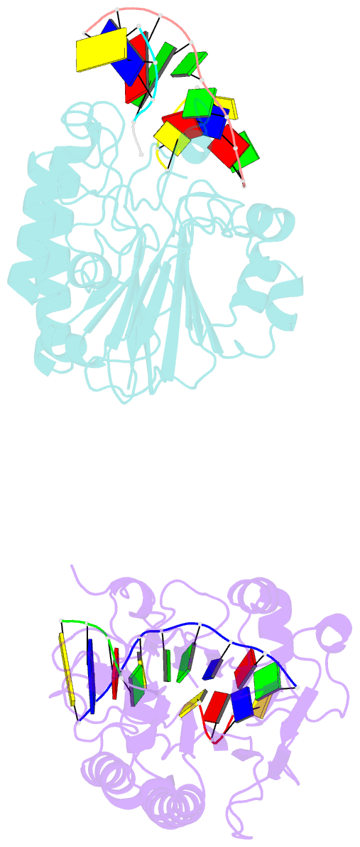
Summary information and primary citation
- PDB-id
- 1de9; SNAP-derived features in text and JSON formats;
DNAproDB
- Class
- lyase-DNA
- Method
- X-ray (3.0 Å)
- Summary
- Human ape1 endonuclease with bound abasic DNA and mn2+ ion
- Reference
- Mol CD, Izumi T, Mitra S, Tainer JA (2000): "DNA-bound structures and mutants reveal abasic DNA binding by APE1 and DNA repair coordination." Nature, 403, 451-456. doi: 10.1038/35000249.
- Abstract
- Non-coding apurinic/apyrimidinic (AP) sites in DNA are continually created in cells both spontaneously and by damage-specific DNA glycosylases. The biologically critical human base excision repair enzyme APE1 cleaves the DNA sugar-phosphate backbone at a position 5' of AP sites to prime DNA repair synthesis. Here we report three co-crystal structures of human APE1 bound to abasic DNA which show that APE1 uses a rigid, pre-formed, positively charged surface to kink the DNA helix and engulf the AP-DNA strand. APE1 inserts loops into both the DNA major and minor grooves and binds a flipped-out AP site in a pocket that excludes DNA bases and racemized beta-anomer AP sites. Both the APE1 active-site geometry and a complex with cleaved AP-DNA and Mn2+ support a testable structure-based catalytic mechanism. Alanine substitutions of the residues that penetrate the DNA helix unexpectedly show that human APE1 is structurally optimized to retain the cleaved DNA product. These structural and mutational results show how APE1 probably displaces bound glycosylases and retains the nicked DNA product, suggesting that APE1 acts in vivo to coordinate the orderly transfer of unstable DNA damage intermediates between the excision and synthesis steps of DNA repair.


