Summary information and primary citation
- PDB-id
- 1dfu; SNAP-derived features in text and JSON formats;
DNAproDB
- Class
- ribosome
- Method
- X-ray (1.8 Å)
- Summary
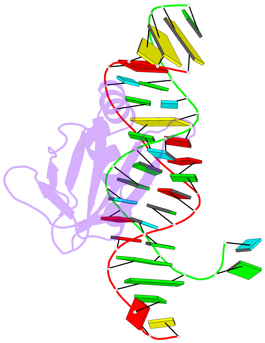
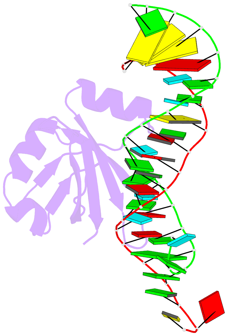
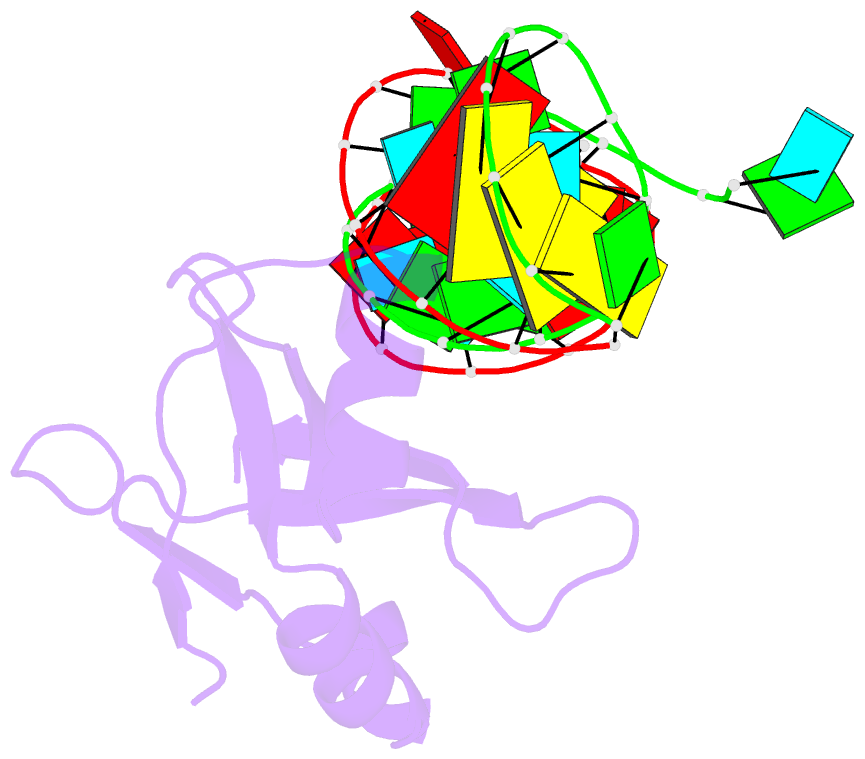
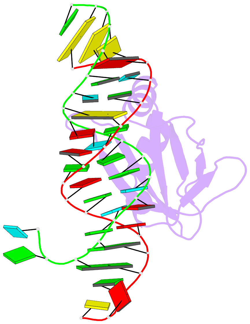
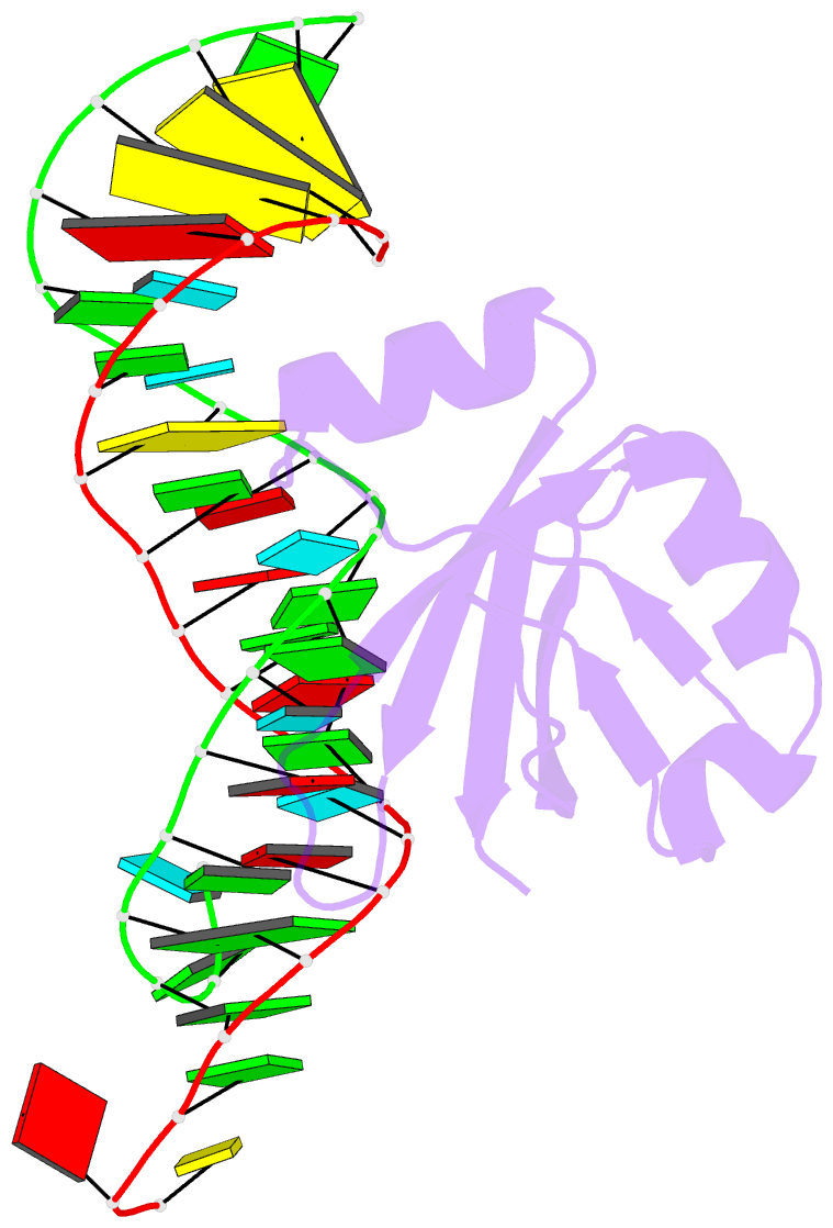
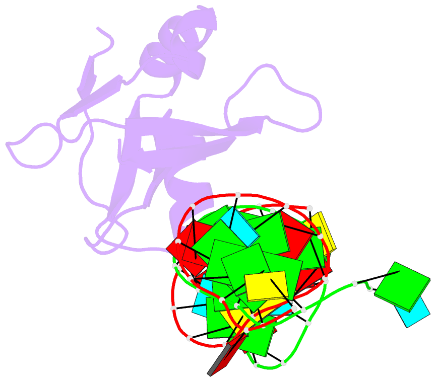
- Crystal structure of e.coli ribosomal protein l25 complexed with a 5s rrna fragment at 1.8 Å resolution
- Reference
- Lu M, Steitz TA (2000): "Structure of Escherichia coli ribosomal protein L25 complexed with a 5S rRNA fragment at 1.8-A resolution." Proc.Natl.Acad.Sci.USA, 97, 2023-2028. doi: 10.1073/pnas.97.5.2023.
- Abstract
- The crystal structure of Escherichia coli ribosomal protein L25 bound to an 18-base pair portion of 5S ribosomal RNA, which contains "loop E," has been determined at 1.8-A resolution. The protein primarily recognizes a unique RNA shape, although five side chains make direct or water-mediated interactions with bases. Three beta-strands lie in the widened minor groove of loop E formed by noncanonical base pairs and cross-strand purine stacks, and an alpha-helix interacts in an adjacent widened major groove. The structure of loop E is largely the same as that of uncomplexed RNA (rms deviation of 0.4 A for 11 base pairs), and 3 Mg(2+) ions that stabilize the noncanonical base pairs lie in the same or similar locations in both structures. Perhaps surprisingly, those residues interacting with the RNA backbone are the most conserved among known L25 sequences, whereas those interacting with the bases are not.