Summary information and primary citation
- PDB-id
- 1di2; SNAP-derived features in text and JSON formats;
DNAproDB
- Class
- RNA binding protein-RNA
- Method
- X-ray (1.9 Å)
- Summary
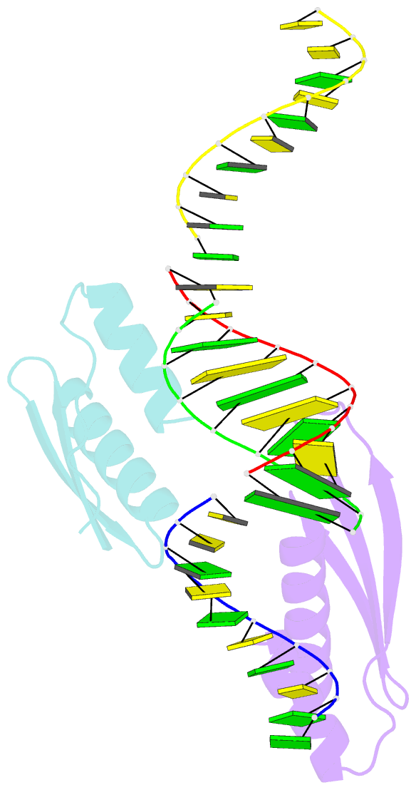
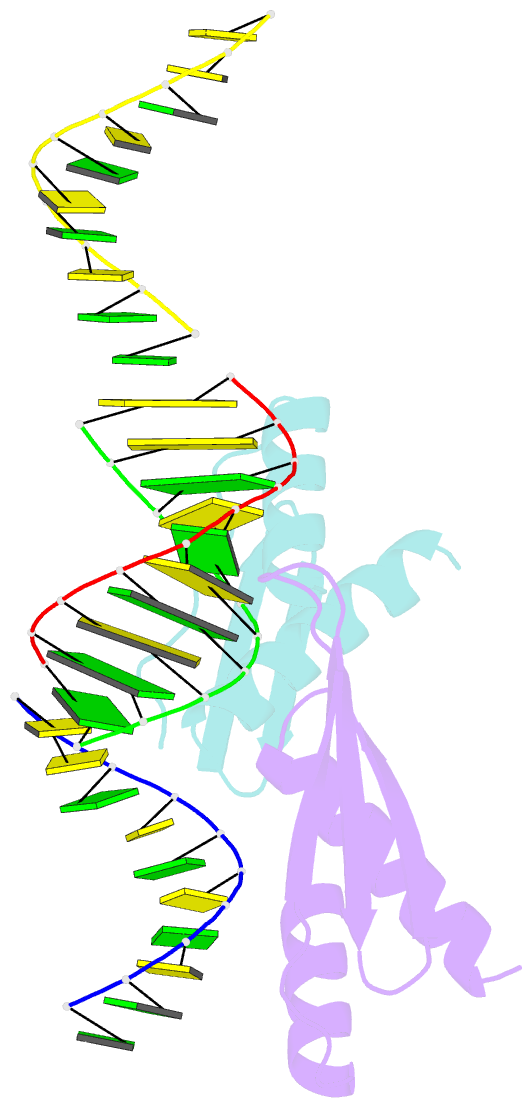
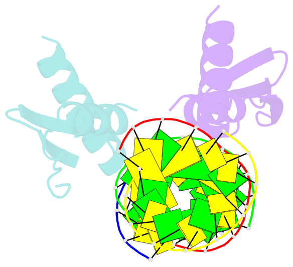
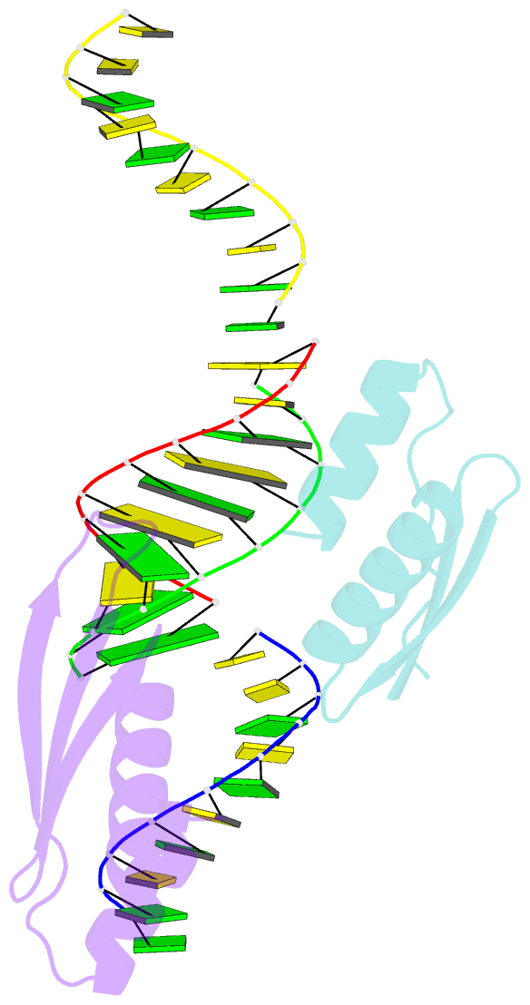
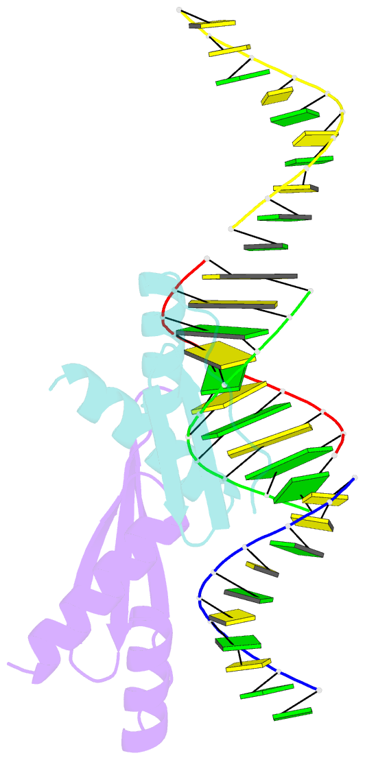
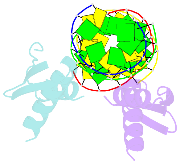
- Crystal structure of a dsrna-binding domain complexed with dsrna: molecular basis of double-stranded RNA-protein interactions
- Reference
- Ryter JM, Schultz SC (1998): "Molecular basis of double-stranded RNA-protein interactions: structure of a dsRNA-binding domain complexed with dsRNA." EMBO J., 17, 7505-7513. doi: 10.1093/emboj/17.24.7505.
- Abstract
- Protein interactions with double-stranded RNA (dsRNA) are critical for many cell processes; however, in contrast to protein-dsDNA interactions, surprisingly little is known about the molecular basis of protein-dsRNA interactions. A large and diverse class of proteins that bind dsRNA do so by utilizing an approximately 70 amino acid motif referred to as the dsRNA-binding domain (dsRBD). We have determined a 1.9 A resolution crystal structure of the second dsRBD of Xenopus laevis RNA-binding protein A complexed with dsRNA. The structure shows that the protein spans 16 bp of dsRNA, interacting with two successive minor grooves and across the intervening major groove on one face of a primarily A-form RNA helix. The nature of these interactions explains dsRBD specificity for dsRNA (over ssRNA or dsDNA) and the apparent lack of sequence specificity. Interestingly, the dsRBD fold resembles a portion of the conserved core structure of a family of polynucleotidyl transferases that includes RuvC, MuA transposase, retroviral integrase and RNase H. Structural comparisons of the dsRBD-dsRNA complex and models proposed for polynucleotidyl transferase-nucleic acid complexes suggest that similarities in nucleic acid binding also exist between these families of proteins.