Summary information and primary citation
- PDB-id
- 1dul; SNAP-derived features in text and JSON formats;
DNAproDB
- Class
- signaling protein-RNA
- Method
- X-ray (1.8 Å)
- Summary
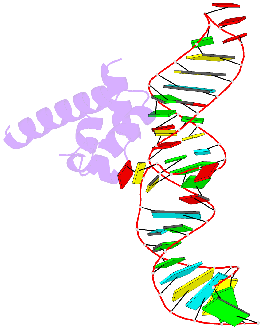
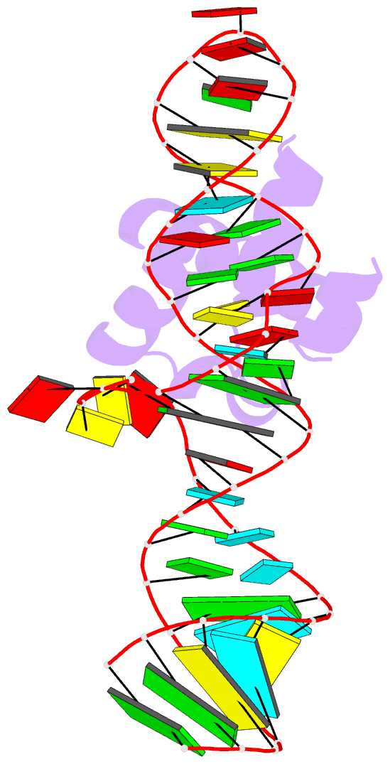
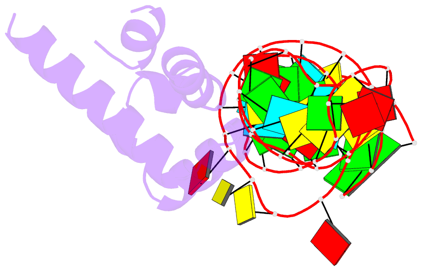
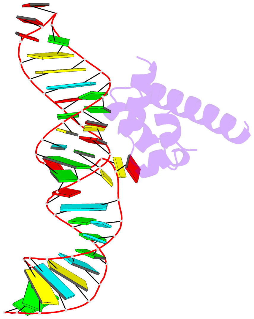
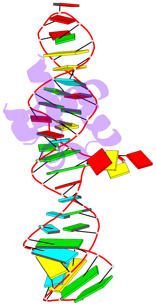
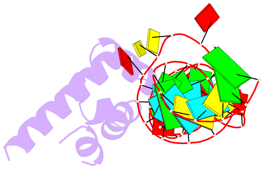
- Structure of the ribonucleoprotein core of the e. coli signal recognition particle
- Reference
- Batey RT, Rambo RP, Lucast L, Rha B, Doudna JA (2000): "Crystal structure of the ribonucleoprotein core of the signal recognition particle." Science, 287, 1232-1239. doi: 10.1126/science.287.5456.1232.
- Abstract
- The signal recognition particle (SRP), a protein-RNA complex conserved in all three kingdoms of life, recognizes and transports specific proteins to cellular membranes for insertion or secretion. We describe here the 1.8 angstrom crystal structure of the universal core of the SRP, revealing protein recognition of a distorted RNA minor groove. Nucleotide analog interference mapping demonstrates the biological importance of observed interactions, and genetic results show that this core is functional in vivo. The structure explains why the conserved residues in the protein and RNA are required for SRP assembly and defines a signal sequence recognition surface composed of both protein and RNA.