Summary information and primary citation
- PDB-id
- 1ea4; SNAP-derived features in text and JSON formats;
DNAproDB
- Class
- gene regulation-DNA
- Method
- X-ray (2.95 Å)
- Summary
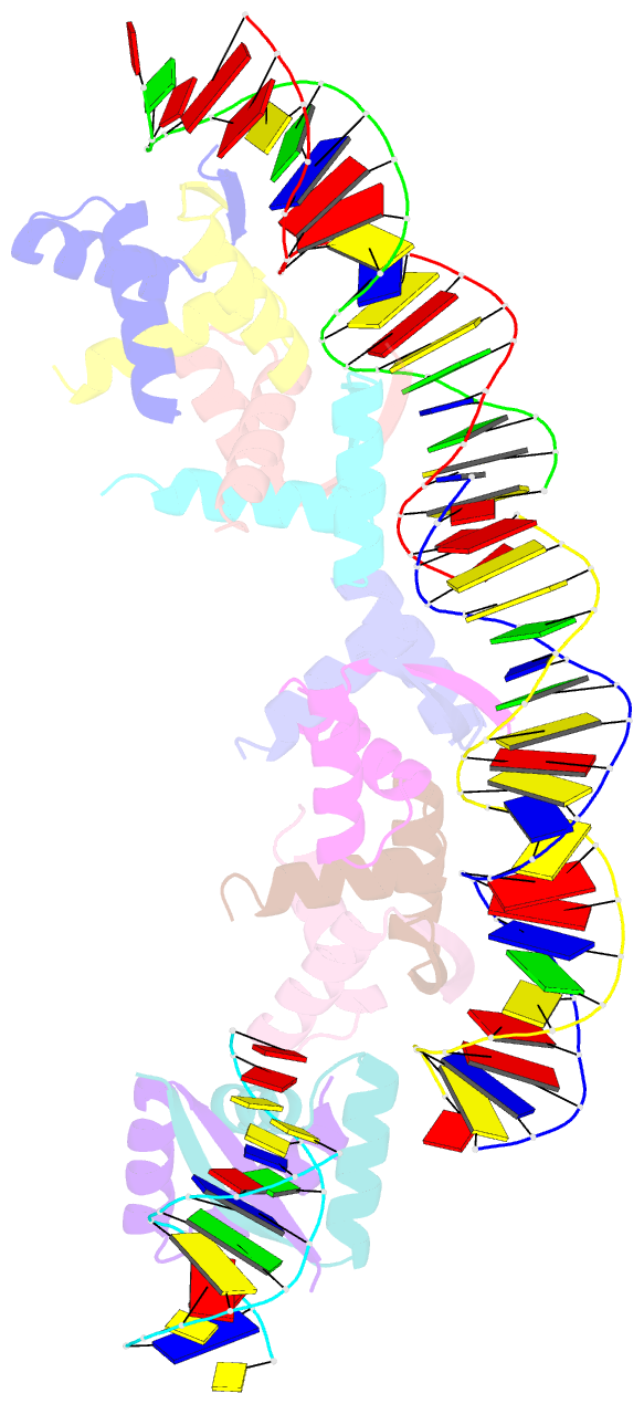
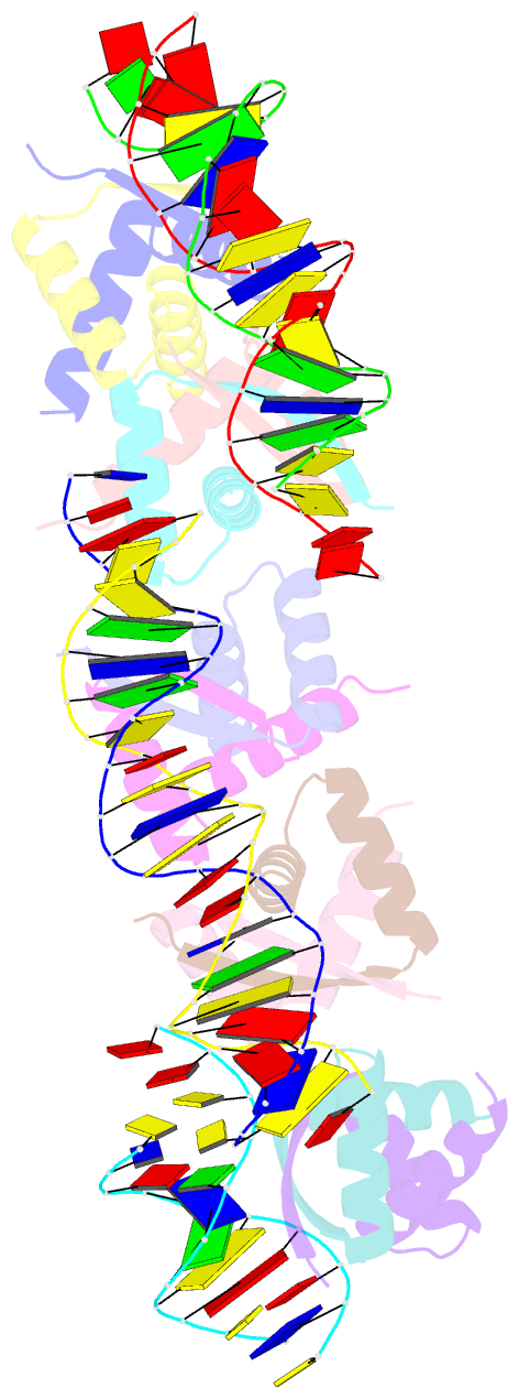
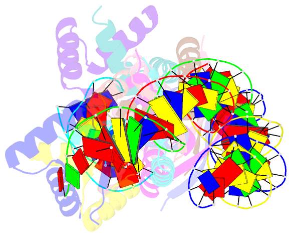
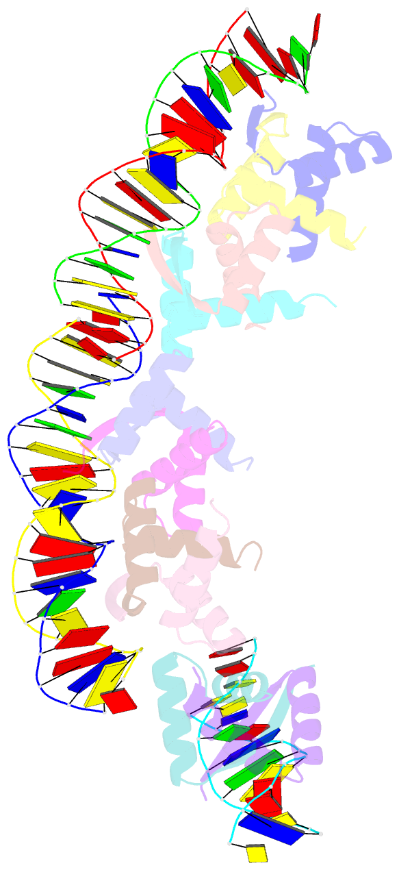
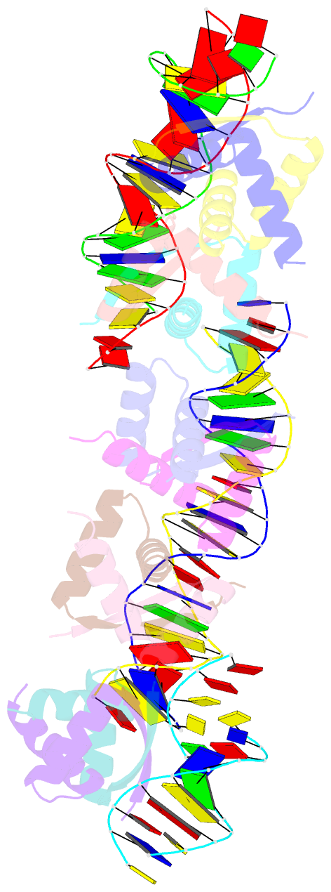
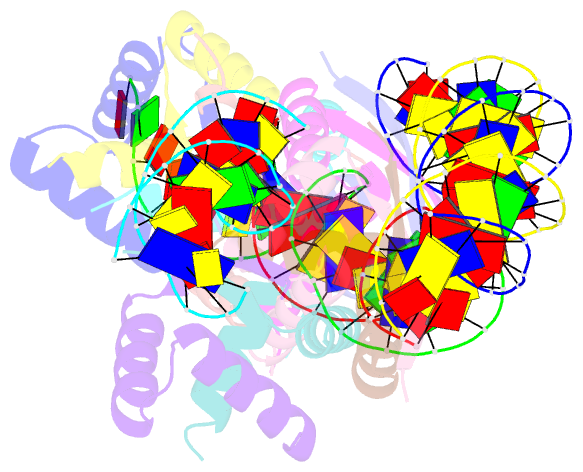
- Transcriptional repressor copg-22bp dsDNA complex
- Reference
- Costa M, Sola M, Del G, Eritja R, Hernaindez-Arriaga AM, Espinosa M, Gomis-Rueth FX, Coll M (2001): "Plasmid Transcriptional Repressor Copg Oligomerises to Render Helical Superstructures Unbound and in Complexes with Oligonucleotides." J.Mol.Biol., 310, 403. doi: 10.1006/JMBI.2001.4760.
- Abstract
- CopG is a 45 amino acid residue transcriptional repressor involved in the copy number control of the streptococcal plasmid pMV158. To do so, it binds to a DNA operator that contains a 13 bp pseudosymmetric DNA element. Binding of CopG to its operator results in repression, at the transcriptional level, of its own synthesis and that of the initiator of replication protein, RepB. Biochemical experiments have shown that CopG co-operatively associates to its target DNA at low protein:DNA ratios, completely protecting four helical turns on the same face of the double helix in both directions from the inverted repeat that constitutes the CopG primary target. This has been correlated with a CopG-mediated DNA bend of about 100 degrees. Here, we show that binding of CopG to DNA fragments containing the inverted repeat just at one end led to nucleation of the protein initiating from the inverted repeat. Nucleation extended to the entire fragment, with CopG-DNA contacts occurring on the same face of the DNA helix. The protein, the prototype for a family of homologous plasmid repressors, displays a homodimeric ribbon-helix-helix arrangement. It polymerises within the unbound crystal to render a continuous right-handed protein superhelix of homodimers, around which a bound double-stranded (ds) DNA could wrap. We have solved the crystal structure of CopG in complex with a 22 bp dsDNA oligonucleotide encompassing the cognate pseudosymmetric element. In the crystal, one protein tetramer binds at one face of the DNA with two parallel beta-ribbons inserted into the major groove. The DNA is bent about 50 degrees under compression of both major and minor grooves. A continuous right-handed complex helix made up mainly by protein-protein and some protein-DNA interactions is observed. The protein-protein interactions involve regions similar to those observed in the oligomerisation of the native crystals and those employed to set up the functional tetramer. A previously solved complex structure of the protein with a 19 bp dsDNA had unveiled a left-handed helical superstructure just made up by DNA interactions.