Summary information and primary citation
- PDB-id
- 1ebm; SNAP-derived features in text and JSON formats;
DNAproDB
- Class
- lyase-DNA
- Method
- X-ray (2.1 Å)
- Summary
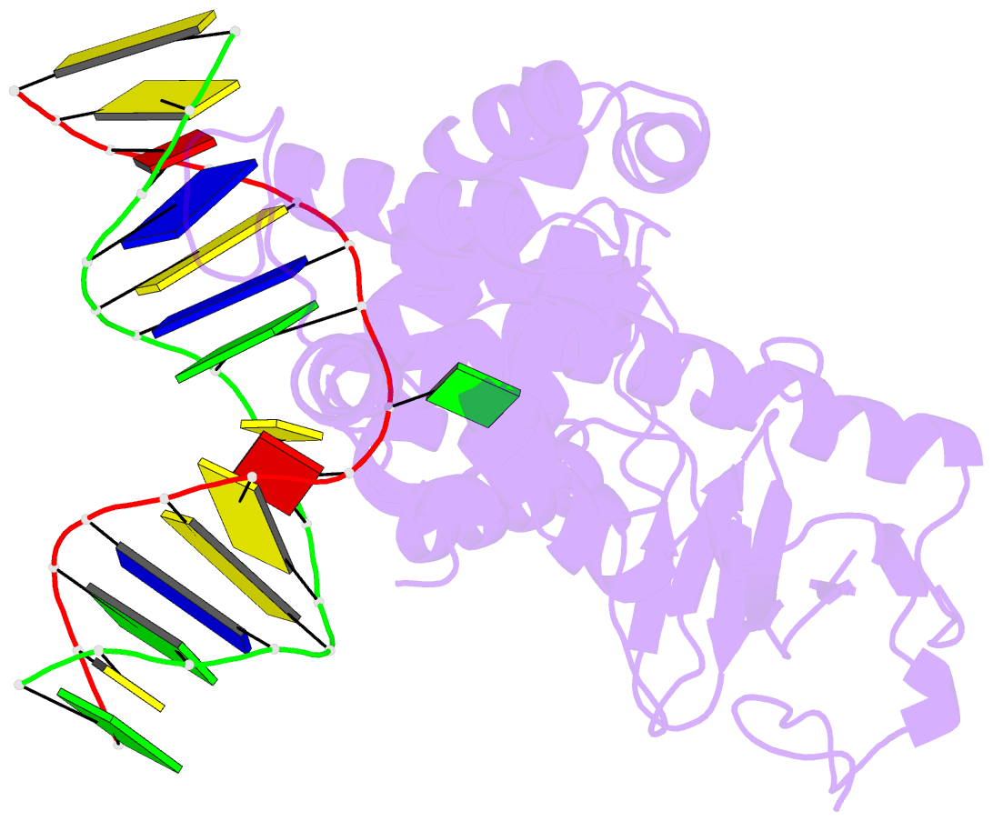
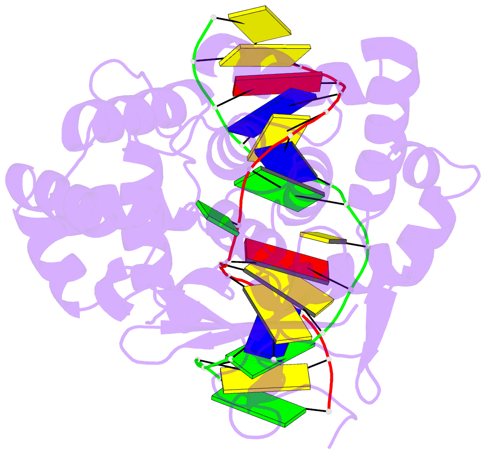
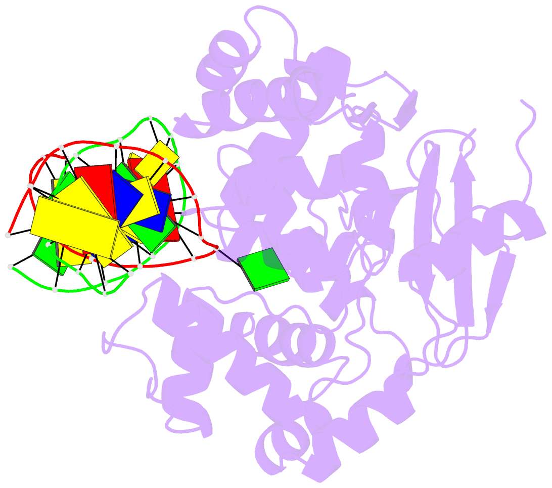
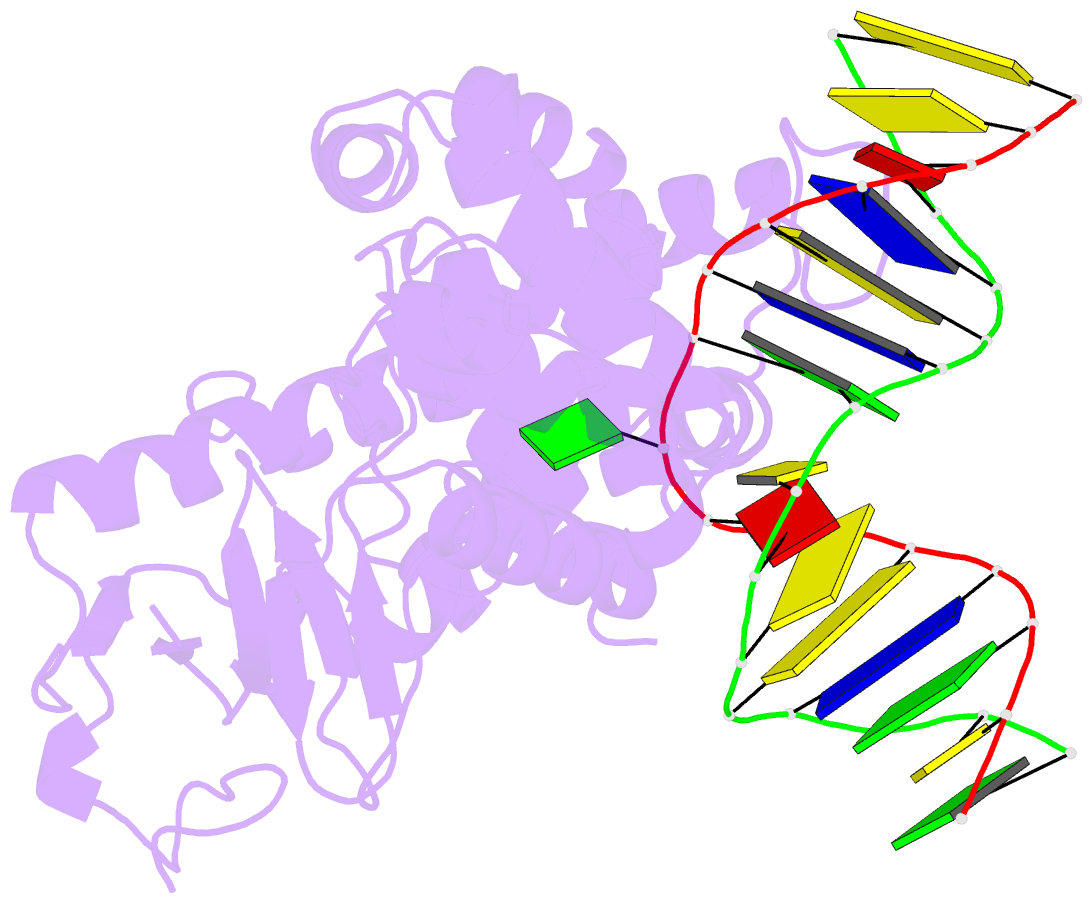
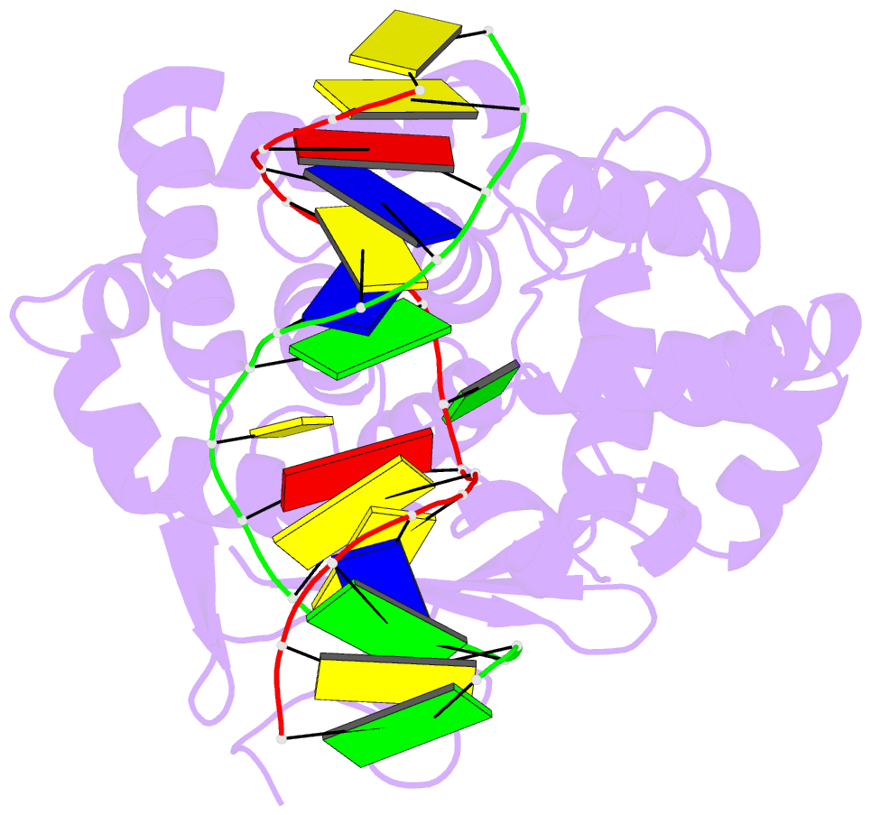
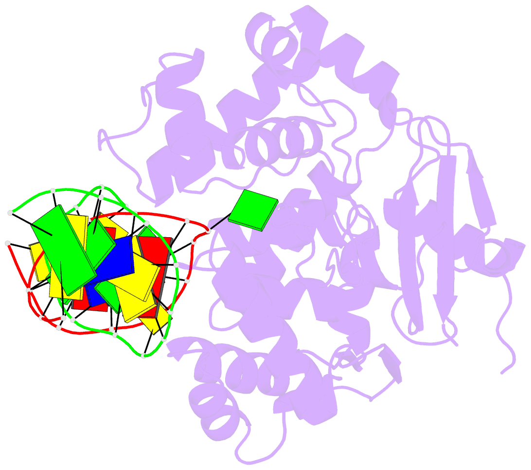
- Crystal structure of the human 8-oxoguanine glycosylase (hogg1) bound to a substrate oligonucleotide
- Reference
- Bruner SD, Norman DP, Verdine GL (2000): "Structural basis for recognition and repair of the endogenous mutagen 8-oxoguanine in DNA." Nature, 403, 859-866. doi: 10.1038/35002510.
- Abstract
- Spontaneous oxidation of guanine residues in DNA generates 8-oxoguanine (oxoG). By mispairing with adenine during replication, oxoG gives rise to a G x C --> T x A transversion, a frequent somatic mutation in human cancers. The dedicated repair pathway for oxoG centres on 8-oxoguanine DNA glycosylase (hOGG1), an enzyme that recognizes oxoG x C base pairs, catalysing expulsion of the oxoG and cleavage of the DNA backbone. Here we report the X-ray structure of the catalytic core of hOGG1 bound to oxoG x C-containing DNA at 2.1 A resolution. The structure reveals the mechanistic basis for the recognition and catalytic excision of DNA damage by hOGG1 and by other members of the enzyme superfamily to which it belongs. The structure also provides a rationale for the biochemical effects of inactivating mutations and polymorphisms in hOGG1. One known mutation, R154H, converts hOGG1 to a promutator by relaxing the specificity of the enzyme for the base opposite oxoG.