Summary information and primary citation
- PDB-id
- 1ehl; SNAP-derived features in text and JSON formats;
DNAproDB
- Class
- immune system
- Method
- X-ray (2.4 Å)
- Summary
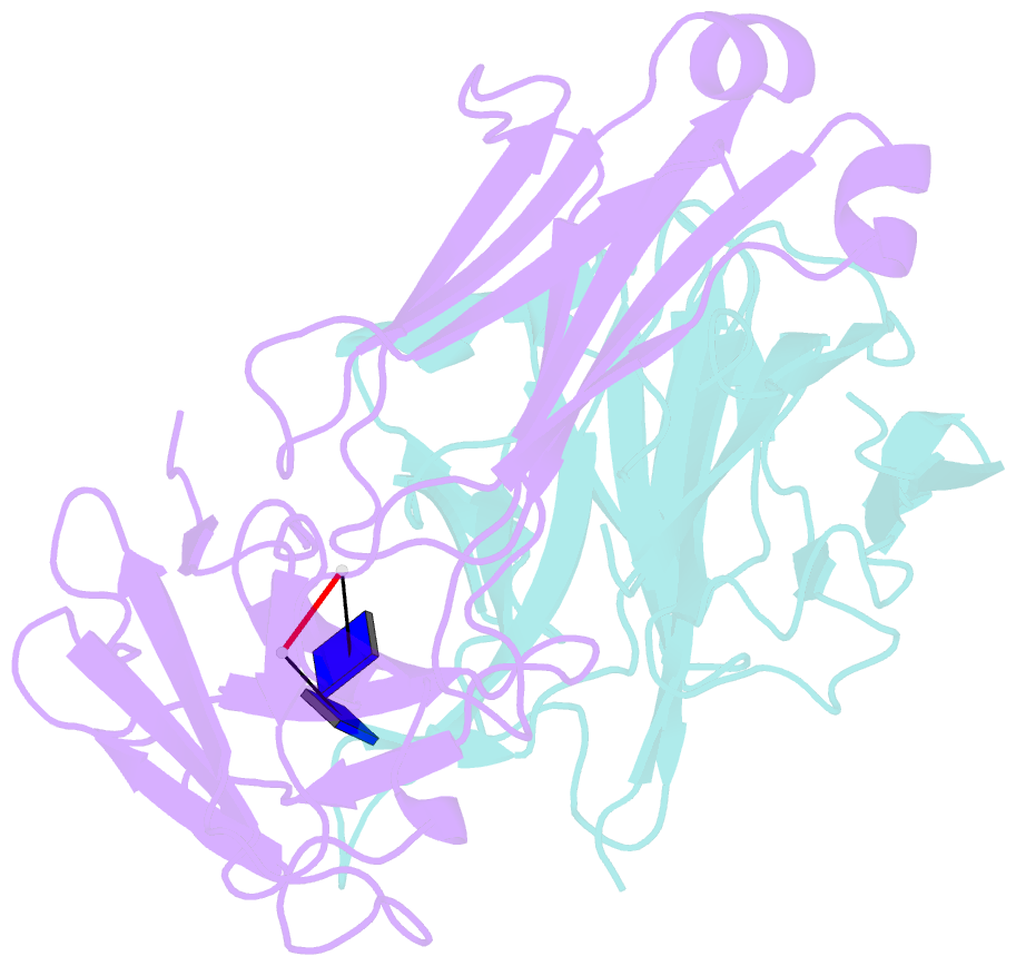
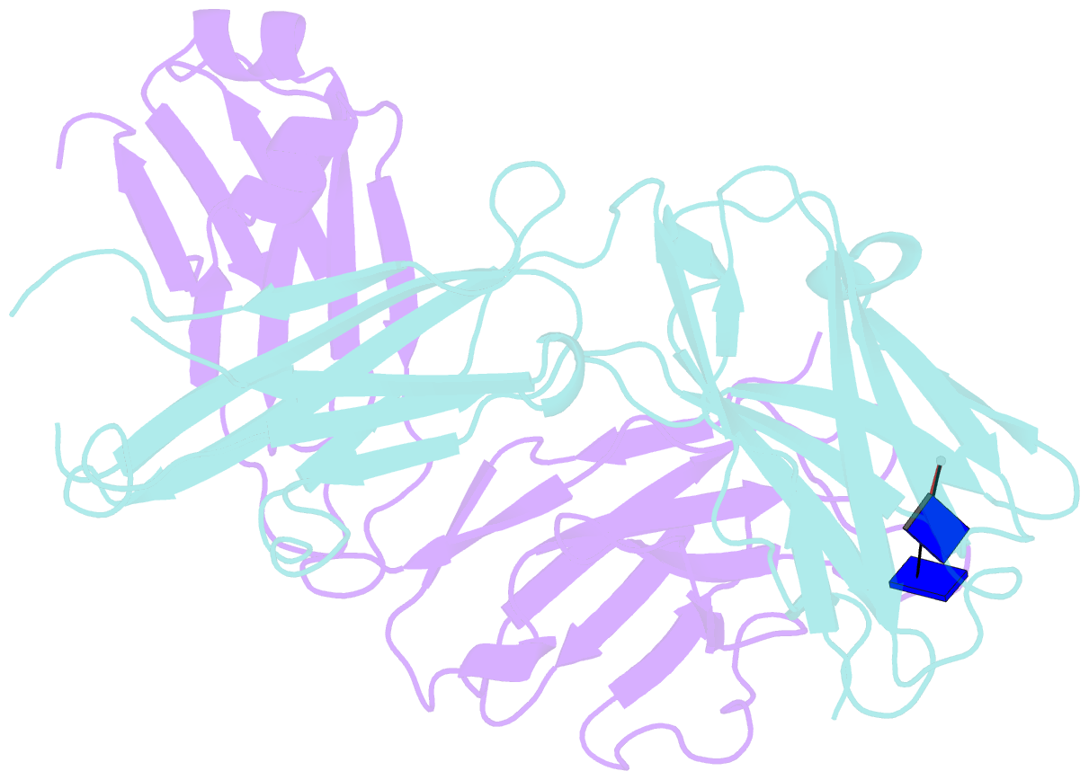
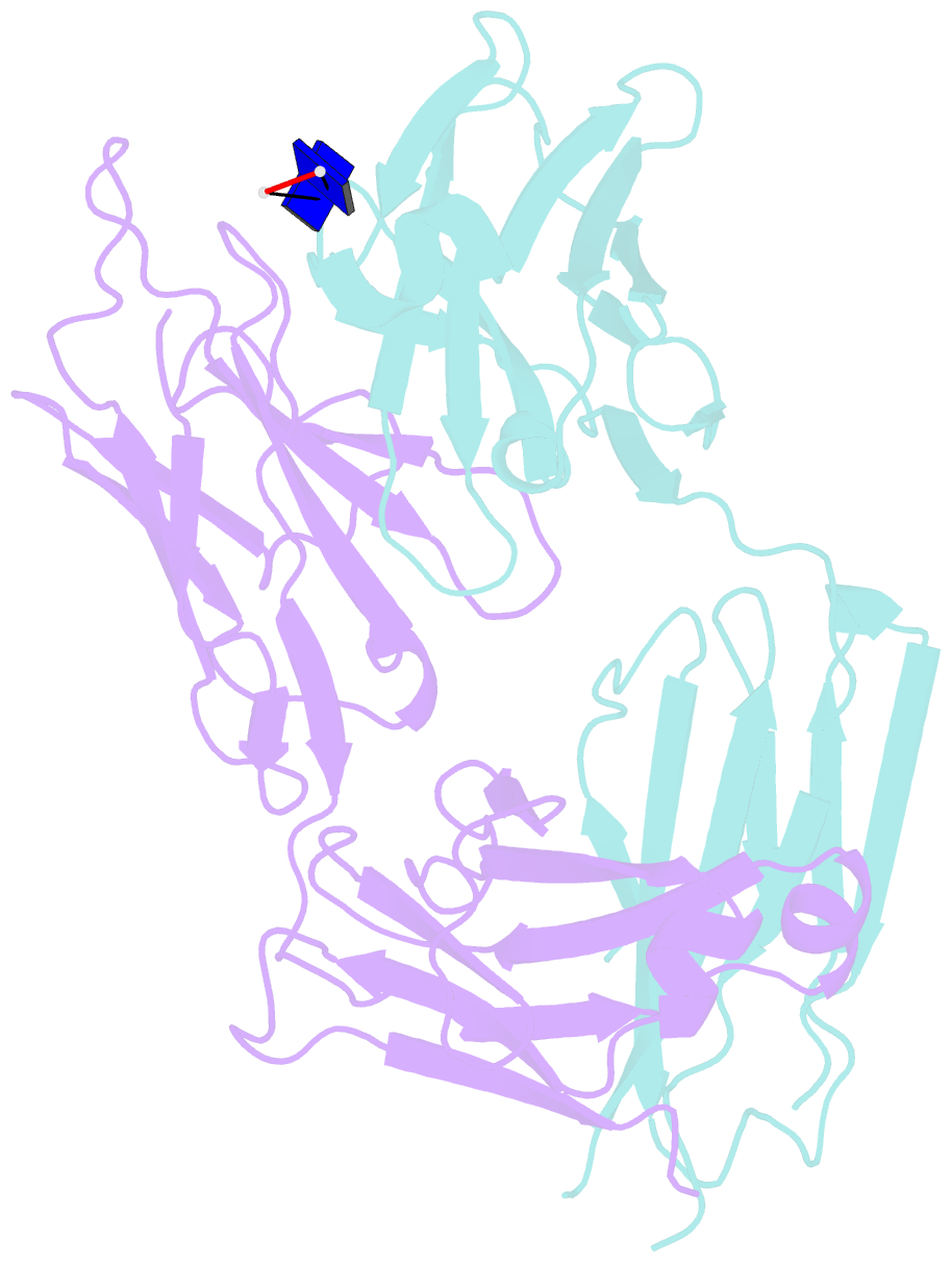
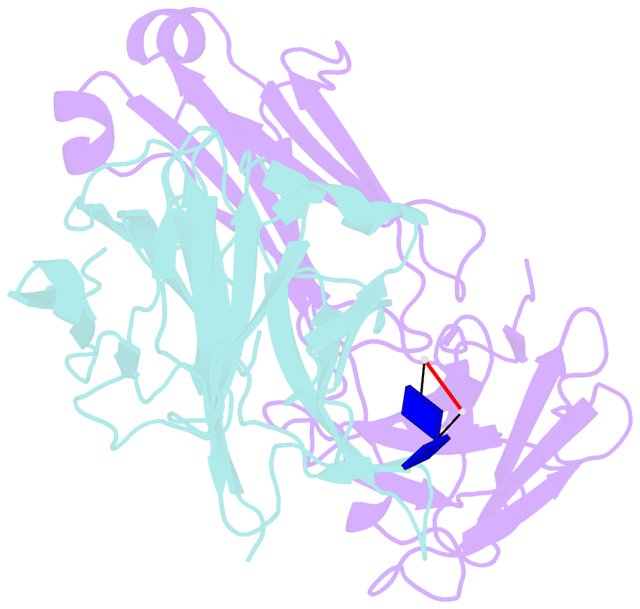
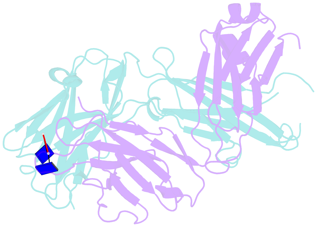
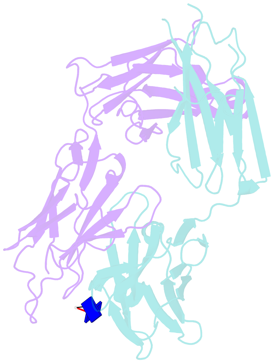
- 64m-2 antibody fab complexed with d(5ht)(6-4)t
- Reference
- Yokoyama H, Mizutani R, Satow Y, Komatsu Y, Ohtsuka E, Nikaido O (2000): "Crystal structure of the 64M-2 antibody Fab fragment in complex with a DNA dT(6-4)T photoproduct formed by ultraviolet radiation." J.Mol.Biol., 299, 711-723. doi: 10.1006/jmbi.2000.3772.
- Abstract
- DNA photoproducts with (6-4) pyrimidine-pyrimidone adducts formed by ultraviolet radiation are implicated in mutagenesis and cancer, particularly skin cancer. The crystal structure of the Fab fragment of the murine 64M-2 antibody specific to DNA T(6-4)T photoproducts is determined as a complex with dT(6-4)T, a (6-4) pyrimidine-pyrimidone photodimer of dTpT, at 2.4 A resolution to a crystallographic R-factor of 0.199 and an R(free) value of 0.279. The 64M-2 Fab molecule is in an extended arrangement with an elbow angle of 174 degrees, and its five complementarity-determining regions, except L2, are involved in the ligand binding. The bound dT(6-4)T ligand adopting a ring structure with (6-4) linked 5' thymine-3' pyrimidone bases is fully accommodated in an antigen-binding pocket of about 15 Ax10 A. The 5'-thymine and 3'-pyrimidone bases are in half-chair and planar conformations, respectively, and are nearly perpendicular to each other. The 5'-thymine base is hydrogen-bonded to Arg95H and Ser96H, and is in van der Waals contact with Tyr100iH. The 3'-pyrimidone base is hydrogen-bonded to His35H, and is in contact with Trp33H. Three water molecules are located at the interface between the bases and the Fab residues. Hydrogen bonds involving these water molecules also contribute to Fab recognition of the dT(6-4)T bases. The sugar-phosphate backbone connecting the bases is surrounded by residues His27dL, Tyr32L, Ser92L, Trp33H, and Ser58H, but is not hydrogen-bonded to these residues.