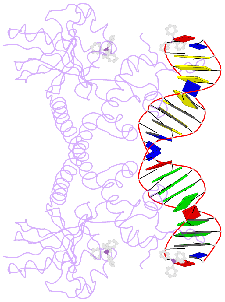
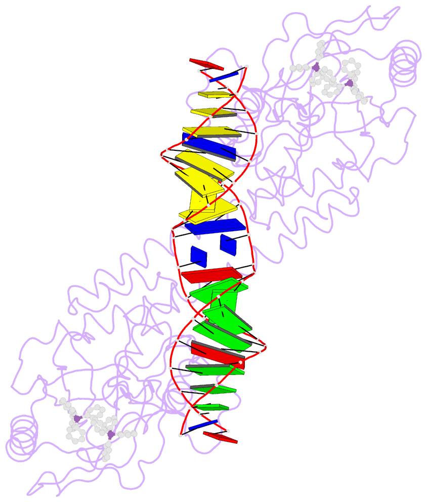
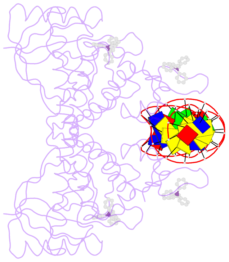
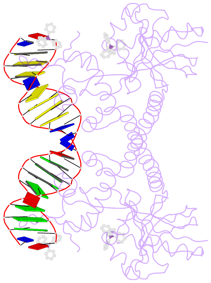
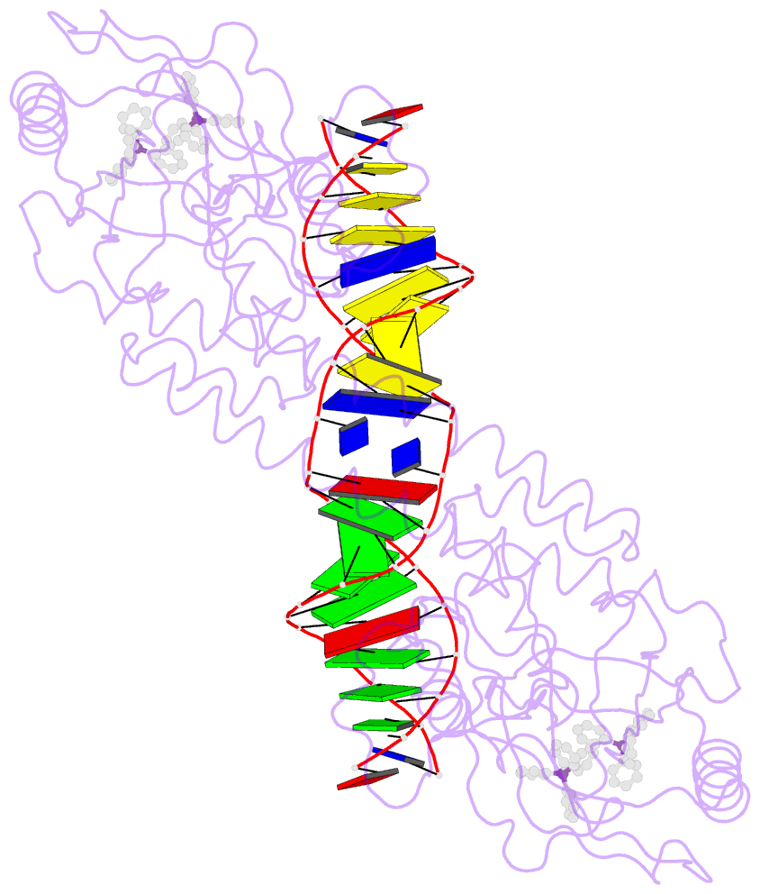
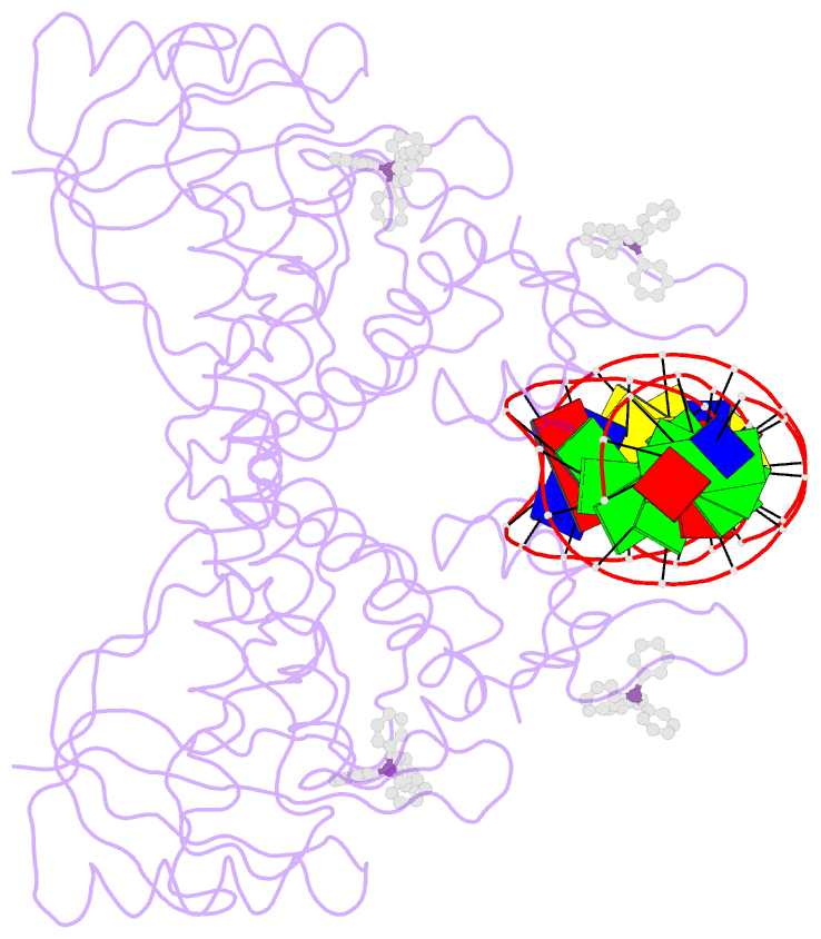
Summary information and primary citation
- PDB-id
- 1exi; SNAP-derived features in text and JSON formats;
DNAproDB
- Class
- transcription-DNA
- Method
- X-ray (3.12 Å)
- Summary
- Crystal structure of transcription activator bmrr, from b. subtilis, bound to 21 base pair bmr operator and tpsb
- Reference
- Heldwein EE, Brennan RG (2001): "Crystal structure of the transcription activator BmrR bound to DNA and a drug." Nature, 409, 378-382. doi: 10.1038/35053138.
- Abstract
- The efflux of chemically diverse drugs by multidrug transporters that span the membrane is one mechanism of multidrug resistance in bacteria. The concentrations of many of these transporters are controlled by transcription regulators, such as BmrR in Bacillus subtilis, EmrR in Escherichia coli and QacR in Staphylococcus aureus. These proteins promote transporter gene expression when they bind toxic compounds. BmrR activates transcription of the multidrug transporter gene, bmr, in response to cellular invasion by certain lipophilic cationic compounds (drugs). BmrR belongs to the MerR family, which regulates response to stress such as exposure to toxic compounds or oxygen radicals in bacteria. MerR proteins have homologous amino-terminal DNA-binding domains but different carboxy-terminal domains, which enable them to bind specific 'coactivator' molecules. When bound to coactivator, MerR proteins upregulate transcription by reconfiguring the 19-base-pair spacer found between the -35 and -10 promoter elements to allow productive interaction with RNA polymerase. Here we report the 3.0 A resolution structure of BmrR in complex with the drug tetraphenylphosphonium (TPP) and a 22-base-pair oligodeoxynucleotide encompassing the bmr promoter. The structure reveals an unexpected mechanism for transcription activation that involves localized base-pair breaking, and base sliding and realignment of the -35 and -10 operator elements.