Summary information and primary citation
- PDB-id
- 1eyg; SNAP-derived features in text and JSON formats;
DNAproDB
- Class
- replication-DNA
- Method
- X-ray (2.8 Å)
- Summary
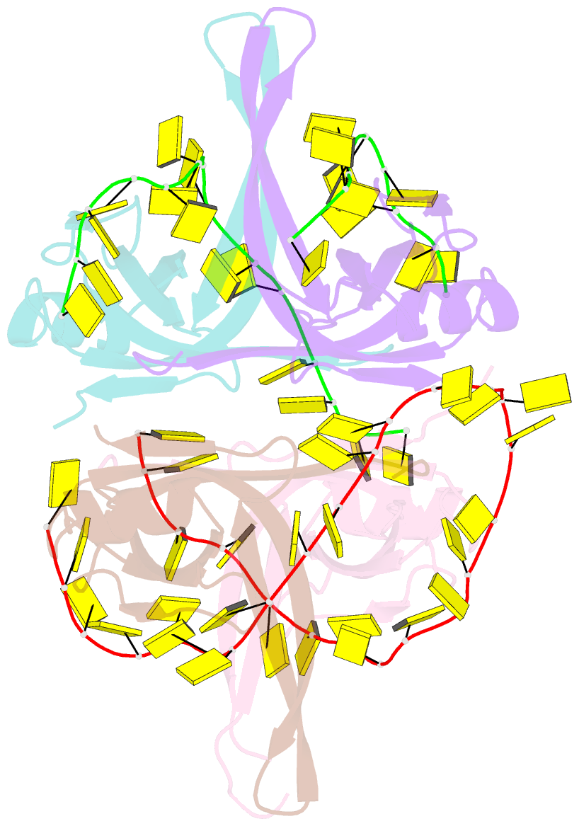
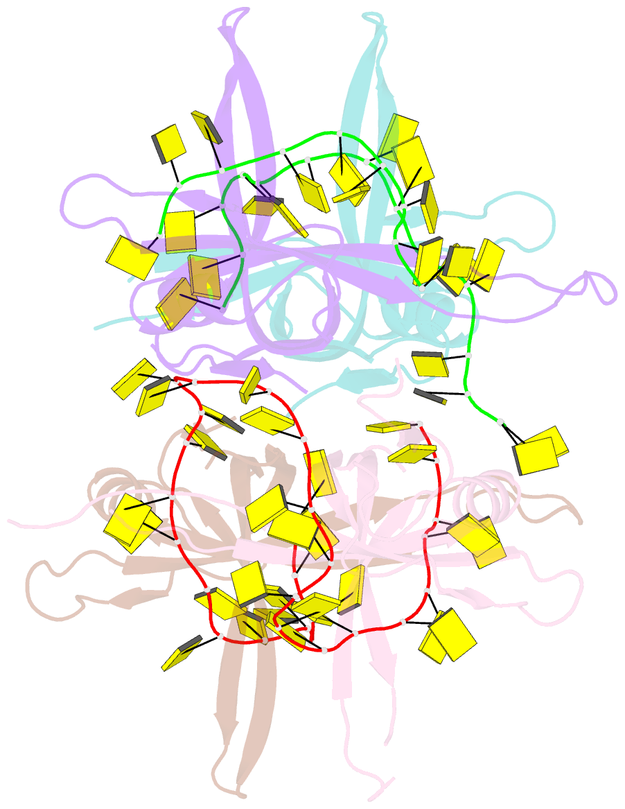
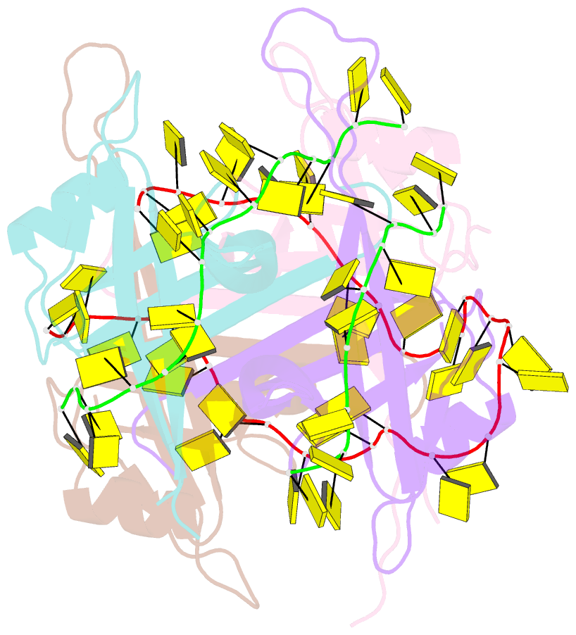
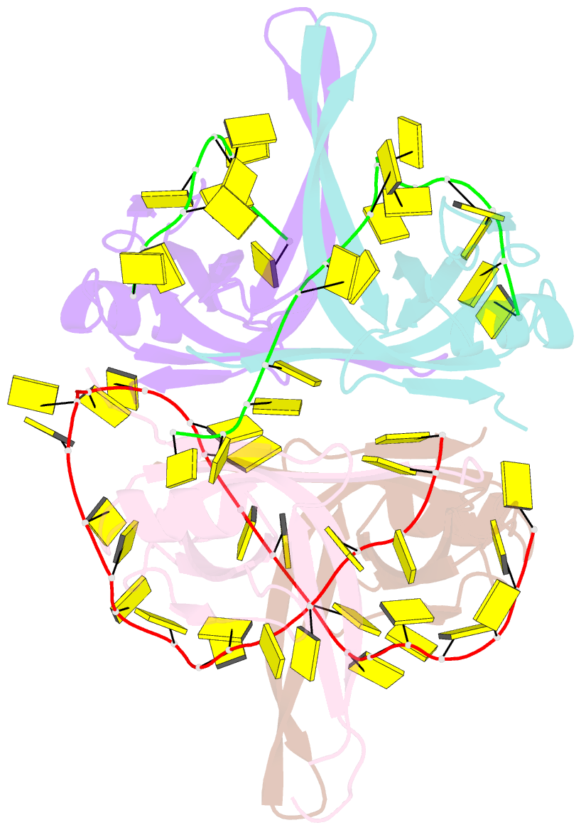
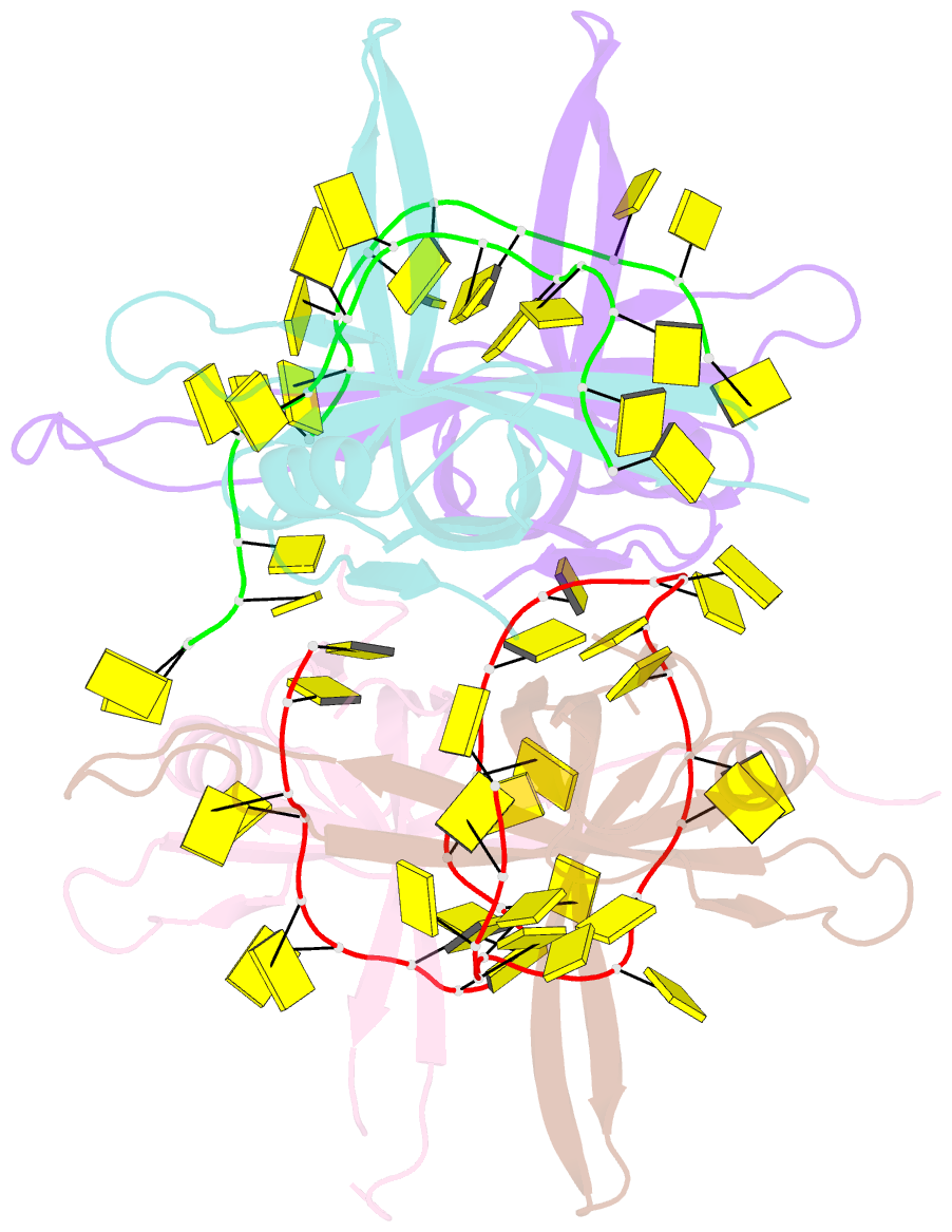
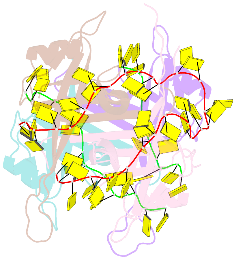
- Crystal structure of chymotryptic fragment of e. coli ssb bound to two 35-mer single strand dnas
- Reference
- Raghunathan S, Kozlov AG, Lohman TM, Waksman G (2000): "Structure of the DNA binding domain of E. coli SSB bound to ssDNA." Nat.Struct.Biol., 7, 648-652. doi: 10.1038/77943.
- Abstract
- The structure of the homotetrameric DNA binding domain of the single stranded DNA binding protein from Escherichia coli (Eco SSB) bound to two 35-mer single stranded DNAs was determined to a resolution of 2.8 A. This structure describes the vast network of interactions that results in the extensive wrapping of single stranded DNA around the SSB tetramer and suggests a structural basis for its various binding modes.