Summary information and primary citation
- PDB-id
- 1f8v; SNAP-derived features in text and JSON formats;
DNAproDB
- Class
- virus-RNA
- Method
- X-ray (3.0 Å)
- Summary
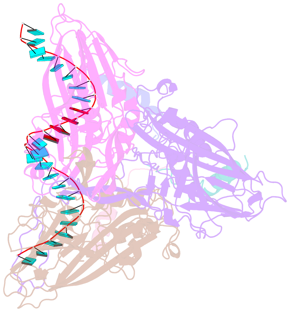
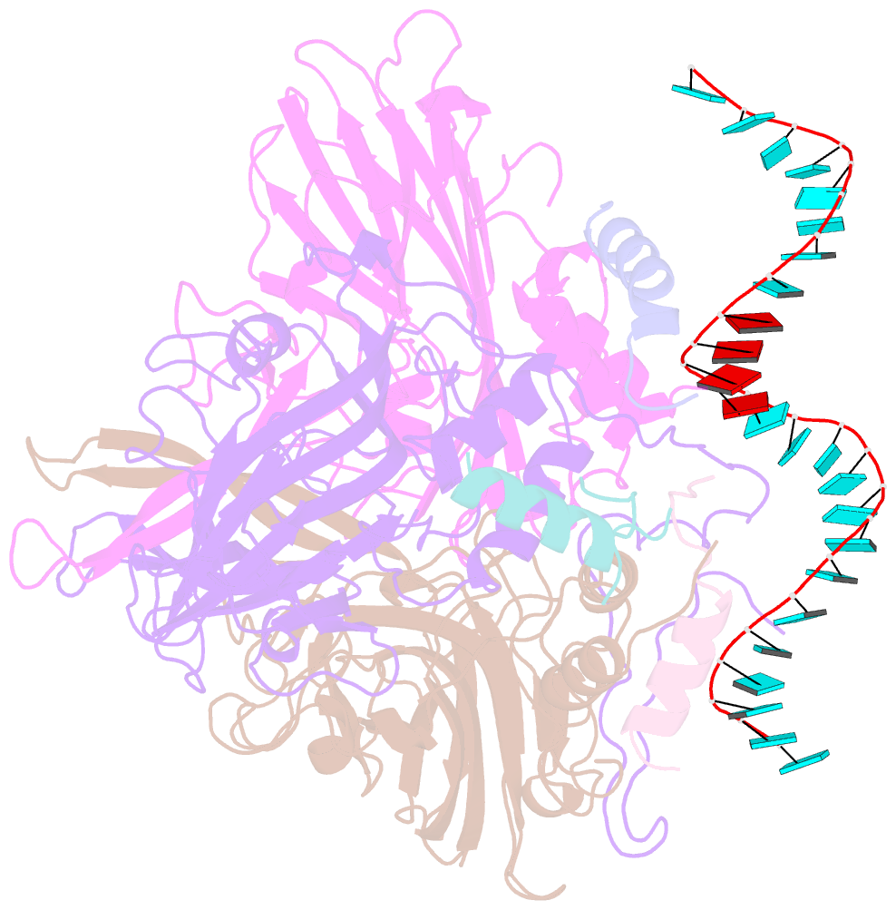
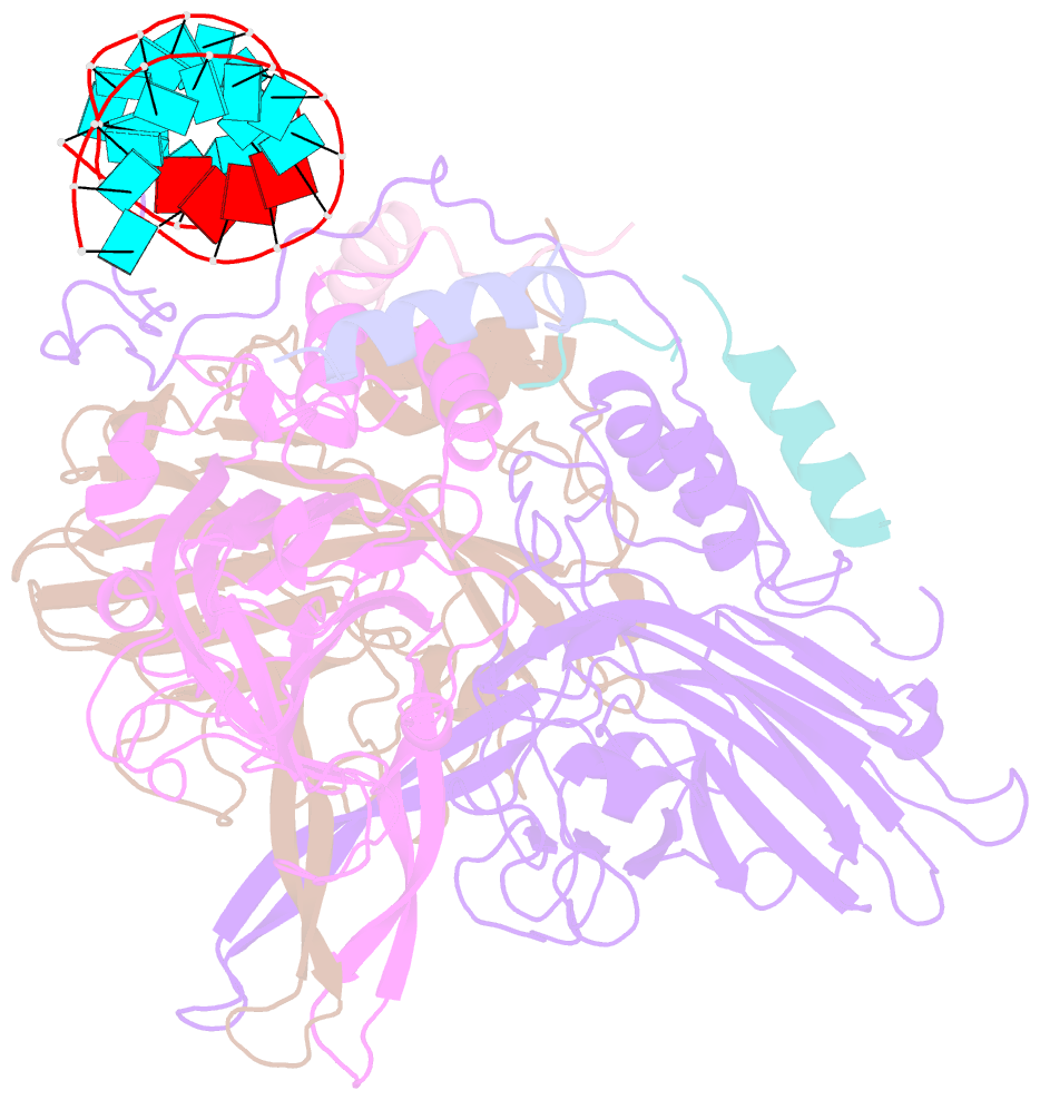
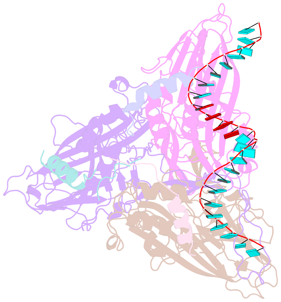
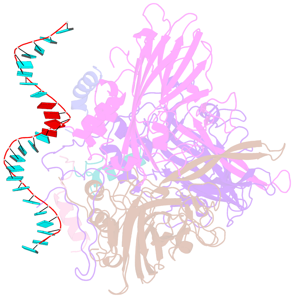
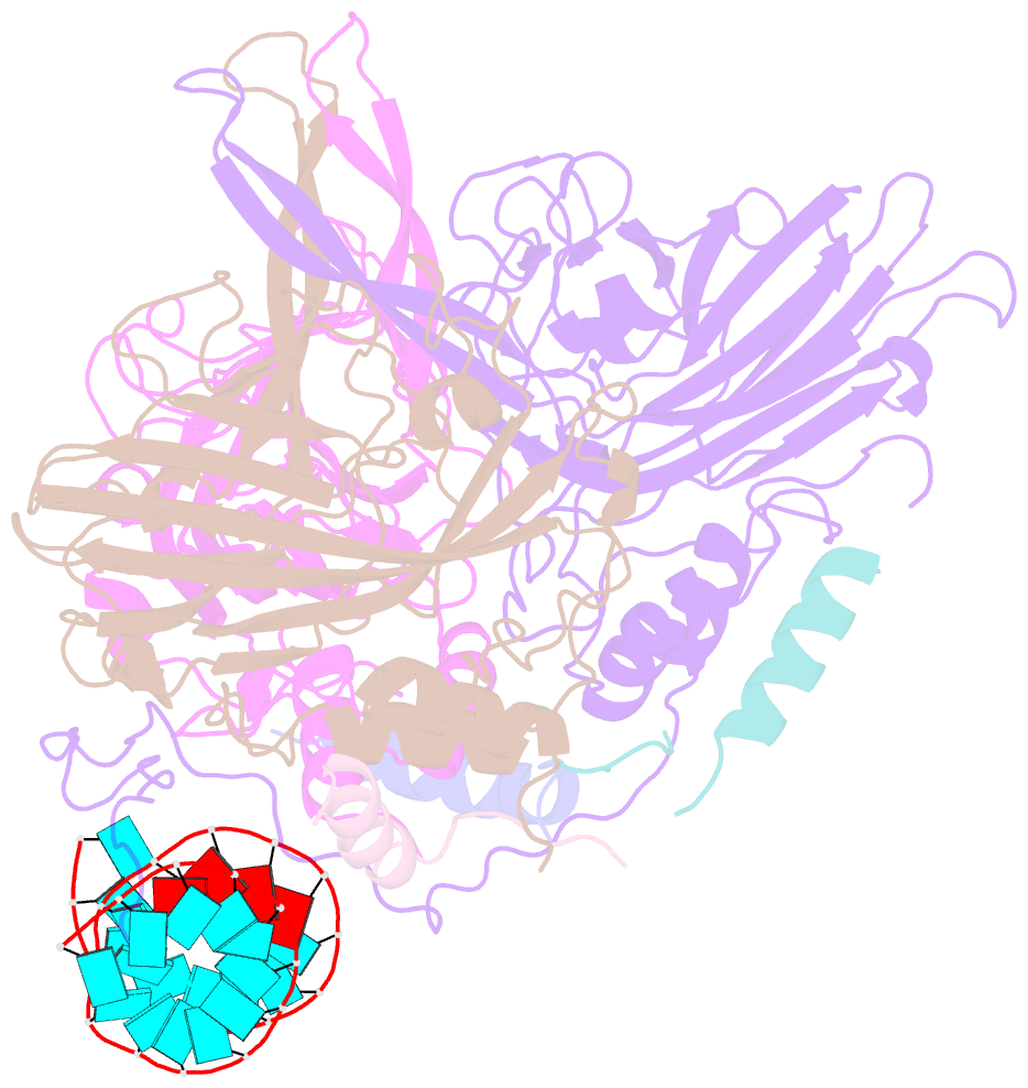
- The structure of pariacoto virus reveals a dodecahedral cage of duplex RNA
- Reference
- Tang L, Johnson KN, Ball LA, Lin T, Yeager M, Johnson JE (2001): "The structure of pariacoto virus reveals a dodecahedral cage of duplex RNA." Nat.Struct.Biol., 8, 77-83. doi: 10.1038/83089.
- Abstract
- The 3.0 A resolution crystal structure of Pariacoto virus (PaV) reveals extensive interactions between portions of the viral RNA genome and the icosahedral capsid. Under the protein shell of the T = 3 quasi equivalent capsid lies a dodecahedral cage composed of RNA duplex that accounts for approximately 35% of the single-stranded RNA genome. The highly basic N-terminal regions (residues 7-54) of the subunits, forming pentamers (A subunits) are clearly visible in the density map and make numerous interactions with the RNA cage. The C-terminal segments (residues 394-401) of the A subunits lie in channels near the quasi three-fold axes. Electron cryo-microscopy and image reconstruction of PaV particles clearly show the dodecahedral RNA cage.