Summary information and primary citation
- PDB-id
- 1fjl; SNAP-derived features in text and JSON formats;
DNAproDB
- Class
- transcription-DNA
- Method
- X-ray (2.0 Å)
- Summary
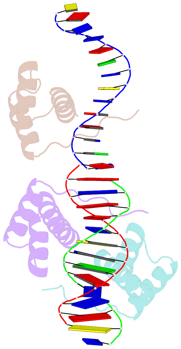
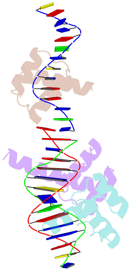
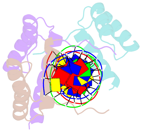
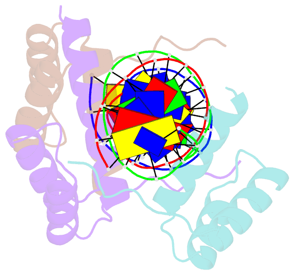
- Homeodomain from the drosophila paired protein bound to a DNA oligonucleotide
- Reference
- Wilson DS, Guenther B, Desplan C, Kuriyan J (1995): "High resolution crystal structure of a paired (Pax) class cooperative homeodomain dimer on DNA." Cell(Cambridge,Mass.), 82, 709-719. doi: 10.1016/0092-8674(95)90468-9.
- Abstract
- The crystal structure of the paired homeodomain bound to DNA as a cooperative dimer has been determined to 2.0 A resolution. Direct contacts between each homeodomain and the DNA are similar to those described previously. In addition, an extensive network of water molecules mediates contacts between the recognition helix and the DNA major groove. Several symmetrical contacts between the two homeodomains underlie the cooperative interaction, and deformations in the DNA structure are necessary for the establishment of these contacts. Comparison with structures of homeodomains bound monomerically to DNA suggests that the binding of a single paired homeodomain can introduce these DNA distortions, thus preparing a template for the cooperative interaction with a second homeodomain. This study shows how the paired (Pax) class homeodomains have achieved cooperativity in DNA binding without the assistance of other domains, thereby enabling the recognition of target sequences that are long enough to ensure specificity.