Summary information and primary citation
- PDB-id
- 1fxl; SNAP-derived features in text and JSON formats;
DNAproDB
- Class
- transcription-RNA
- Method
- X-ray (1.8 Å)
- Summary
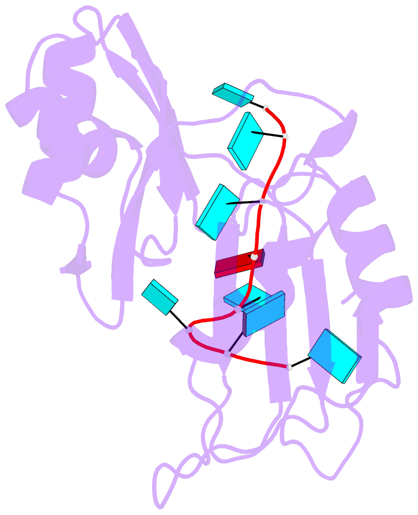
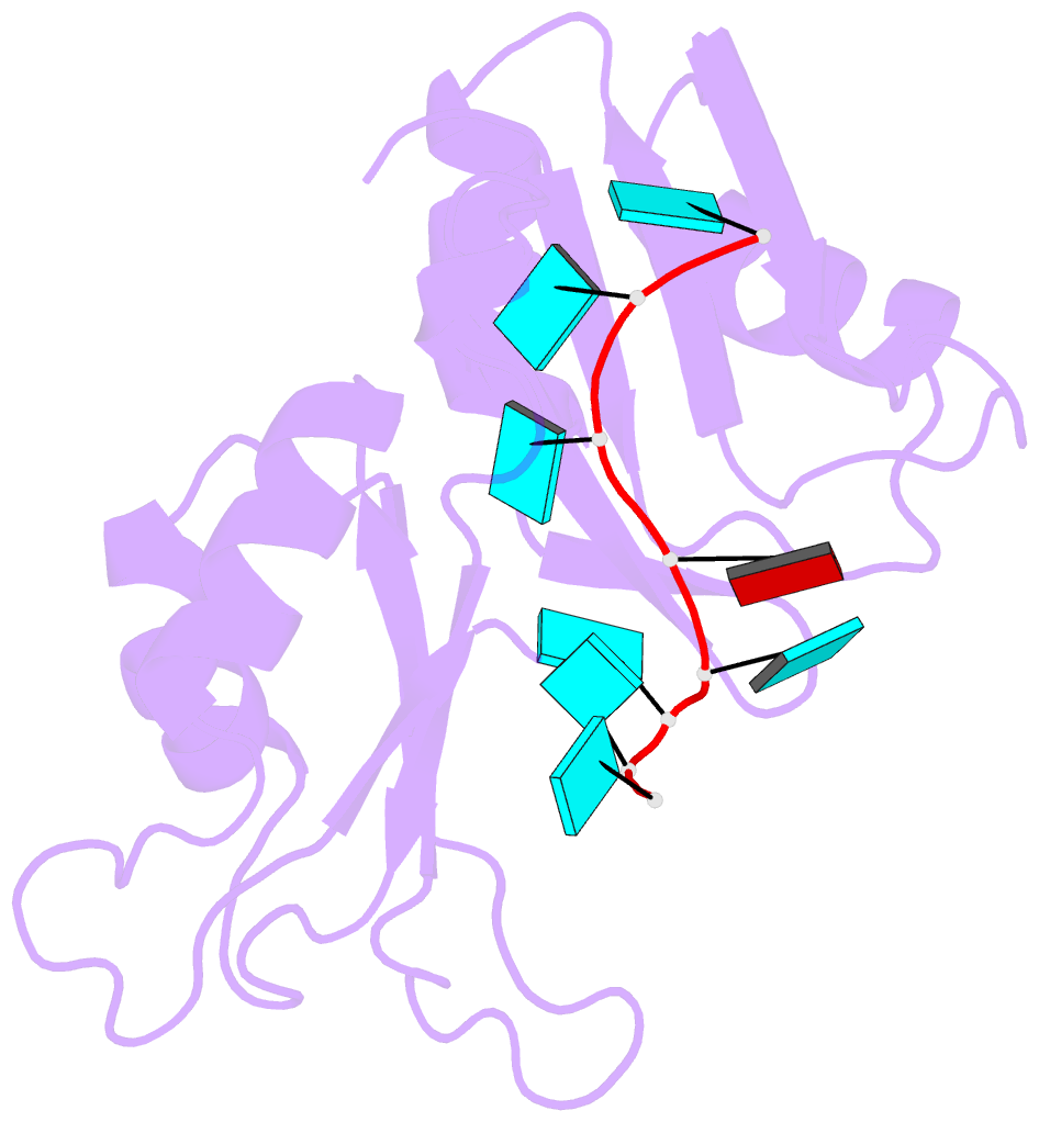
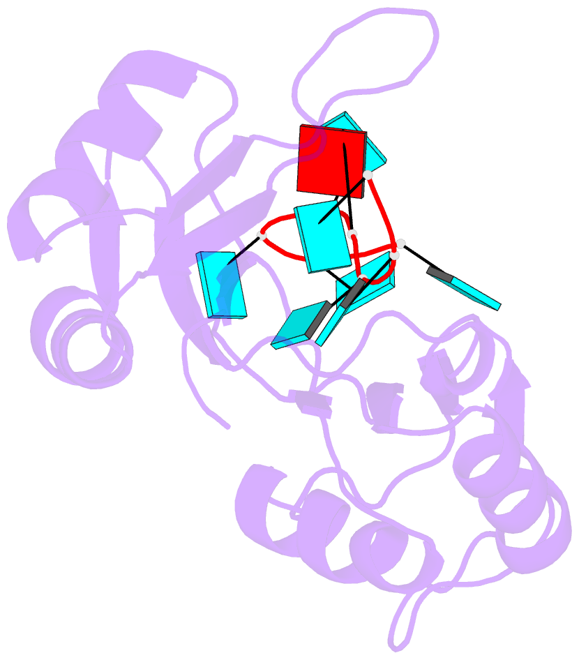
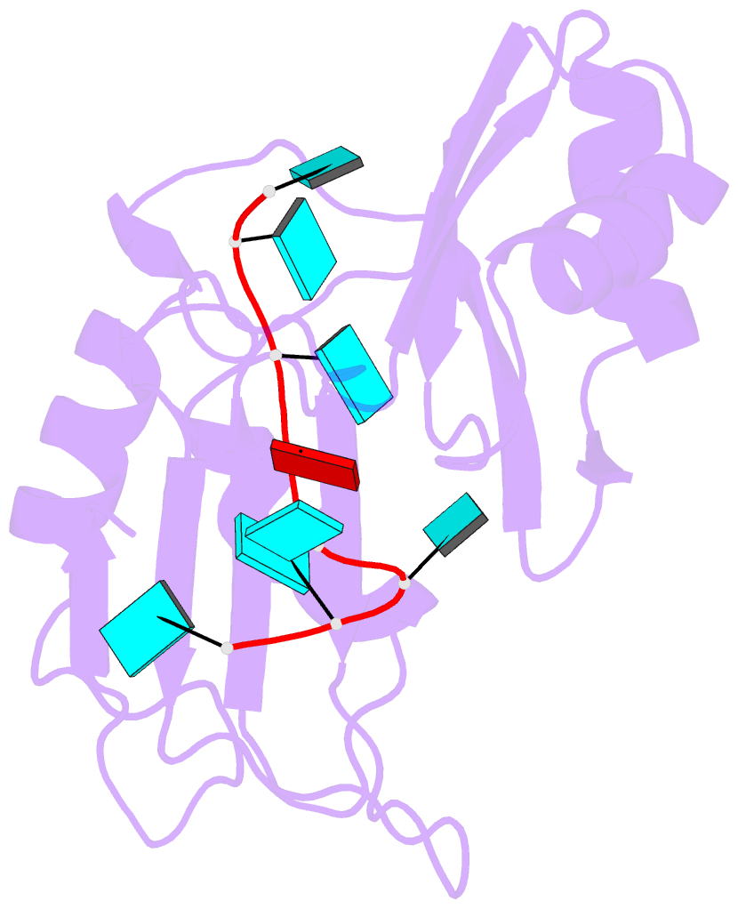
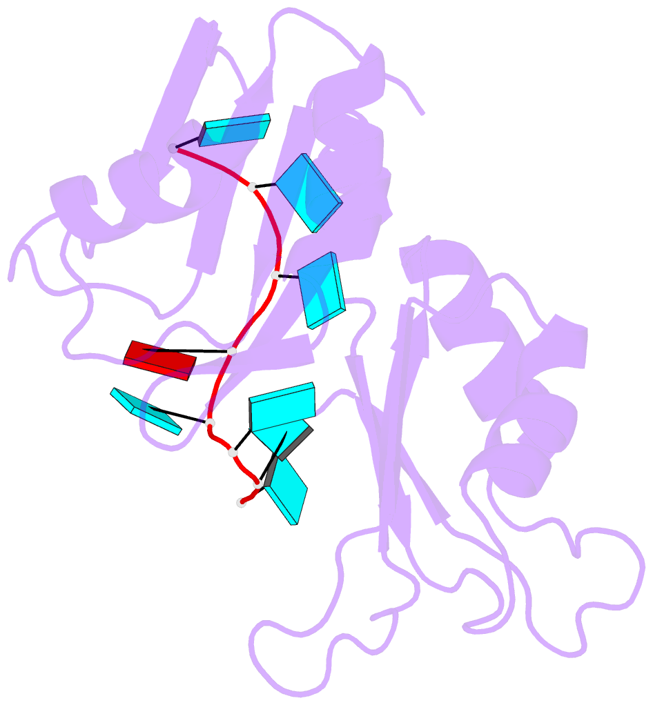
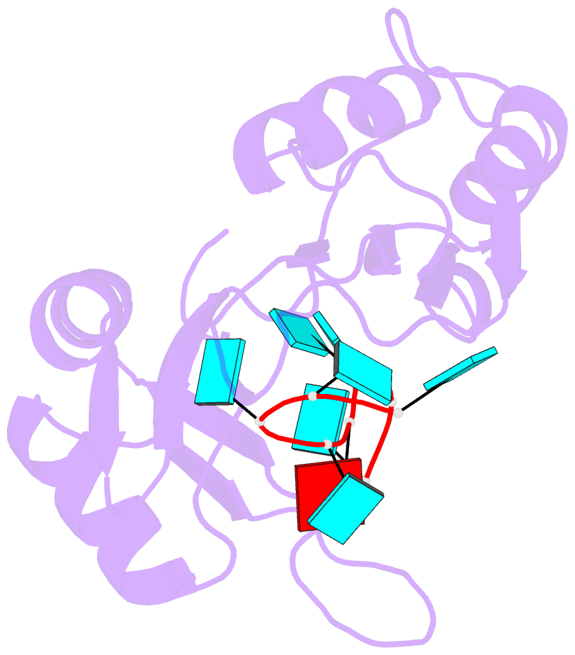
- Crystal structure of hud and au-rich element of the c-fos RNA
- Reference
- Wang X, Tanaka Hall TM (2001): "Structural basis for recognition of AU-rich element RNA by the HuD protein." Nat.Struct.Biol., 8, 141-145. doi: 10.1038/84131.
- Abstract
- Hu proteins bind to adenosine-uridine (AU)-rich elements (AREs) in the 3' untranslated regions of many short-lived mRNAs, thereby stabilizing them. Here we report the crystal structures of the first two RNA recognition motif (RRM) domains of the HuD protein in complex with an 11-nucleotide fragment of a class I ARE (the c-fos ARE; to 1.8 A), and with an 11-nucleotide fragment of a class II ARE (the tumor necrosis factor alpha ARE; to 2.3 A). These structures reveal a consensus RNA recognition sequence that suggests a preference for pyrimidine-rich sequences and a requirement for a central uracil residue in the clustered AUUUA repeats found in class II AREs. Comparison to structures of other RRM domain-nucleic acid complexes reveals two base recognition pockets in all the structures that interact with bases using residues in conserved ribonucleoprotein motifs and at the C-terminal ends of RRM domains. Different conformations of nucleic acid can be bound by RRM domains by using different combinations of base recognition pockets and multiple RRM domains.