Summary information and primary citation
- PDB-id
- 1g3x; SNAP-derived features in text and JSON formats;
DNAproDB
- Class
- DNA
- Method
- X-ray (2.7 Å)
- Summary
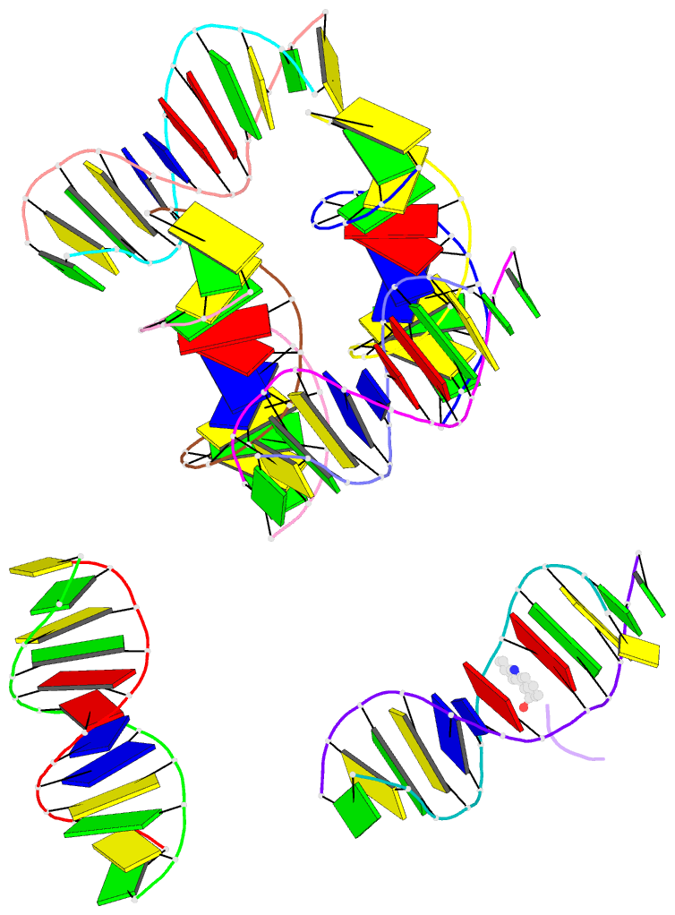
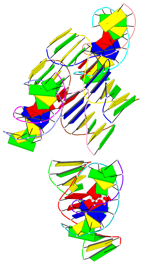
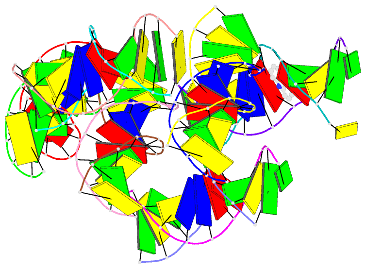
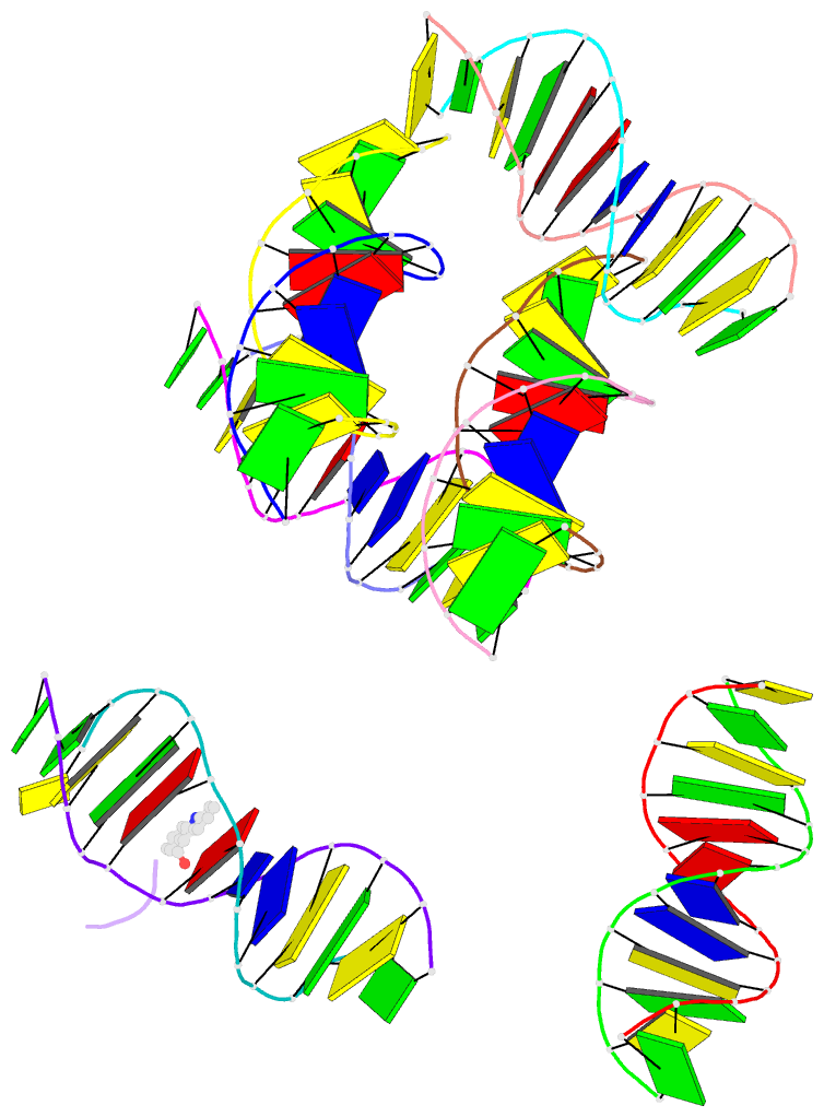
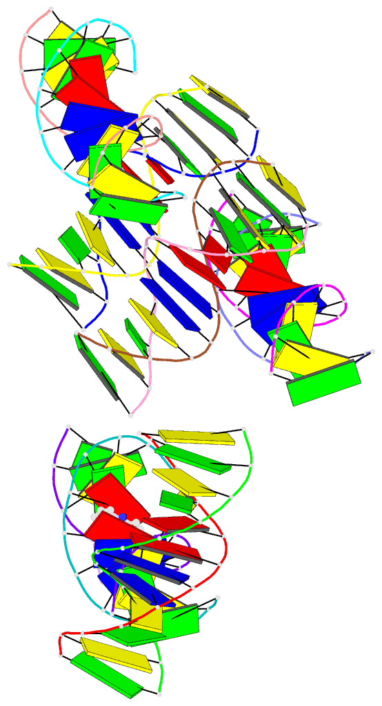
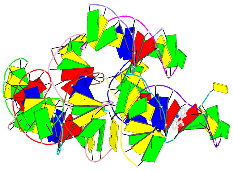
- Intercalation of an 9acridine-peptide drug in a DNA dodecamer
- Reference
- Malinina L, Soler-Lopez M, Aymami J, Subirana JA (2002): "Intercalation of an acridine-peptide drug in an AA/TT base step in the crystal structure of [d(CGCGAATTCGCG)](2) with six duplexes and seven Mg(2+) ions in the asymmetric unit." Biochemistry, 41, 9341-9348. doi: 10.1021/bi020135c.
- Abstract
- We present the crystal structure of an acridine drug derivatized at carbon 9, [N(alpha)-(9-acridinoyl)-tetraarginine], intercalated within the dodecamer [d(CGCGAATTCGCG)](2). The presence of a lateral chain at the central carbon 9 atom differentiates this compound from most acridine drugs hitherto studied, which are usually derivatized at carbon 4. The DNA:drug interaction we observe differs from that observed in previous studies, which primarily involves shorter, mainly hexameric sequences, in two important regards: the acridine intercalates within an AA/TT base step, rather than within a CG/CG base step; and the binding site is located at the center of the sequence, rather than at one end of the duplex. In addition, we observe a novel crystal packing arrangement, with six dodecamer duplexes and seven hydrated magnesium ions in the asymmetric unit of a large (66.5 x 68.4 x 77.4 A(3)) unit cell in space group P2(1)2(1)2(1). The duplexes are organized in layers parallel to the ab plane, with consecutive layers crossing each other at right angles.