Summary information and primary citation
- PDB-id
- 1g4d; SNAP-derived features in text and JSON formats;
DNAproDB
- Class
- viral protein-DNA
- Method
- NMR
- Summary
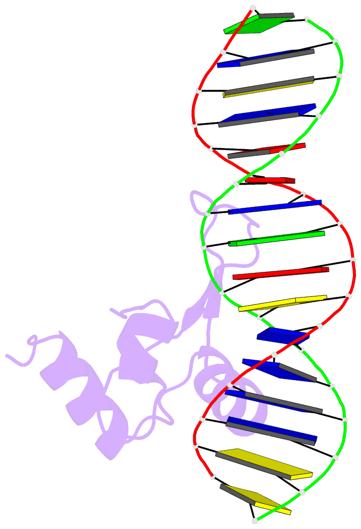
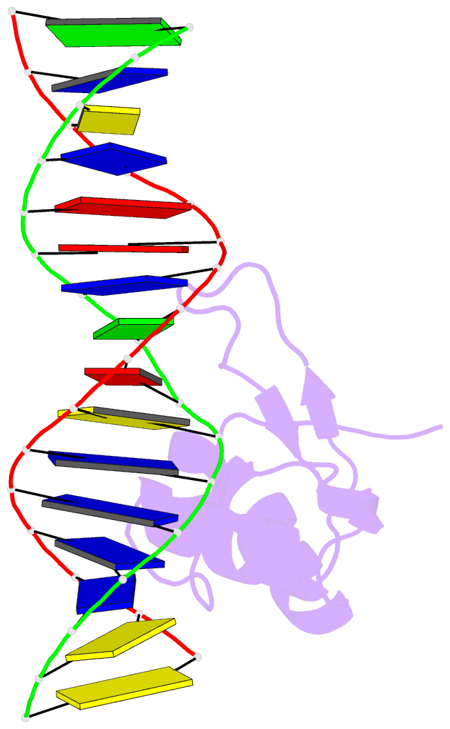
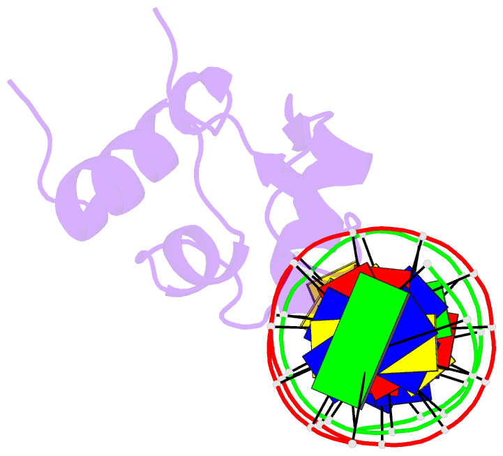
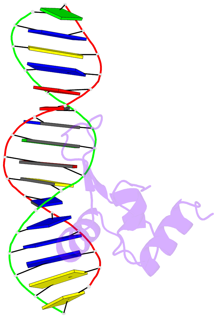
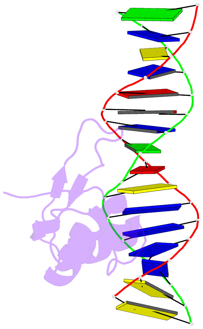
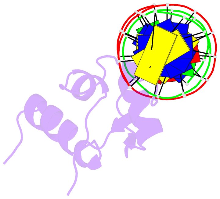
- NMR structure of the mu bacteriophage repressor DNA-binding domain-DNA complex
- Reference
- Wojciak JM, Iwahara J, Clubb RT (2001): "The Mu repressor-DNA complex contains an immobilized 'wing' within the minor groove." Nat.Struct.Biol., 8, 84-90. doi: 10.1038/89582.
- Abstract
- We have determined the solution structure of the complex between the 'winged-helix' enhancer binding domain of the Mu repressor protein and its cognate DNA site. The structure reveals an unusual use for the 'wing' which becomes immobilized upon DNA binding where it makes intermolecular hydrogen bond contacts deep within the minor groove. Although the wing is mobile in the absence of DNA, it partially negates the large entropic penalty associated with its burial by maintaining a small degree of structural order in the DNA-free state. Extensive contacts are also formed between the recognition helix and the DNA, which reads the major groove of a highly conserved region of the binding site through a single base-specific hydrogen bond and van der Waals contacts.