Summary information and primary citation
- PDB-id
- 1gcc; SNAP-derived features in text and JSON formats;
DNAproDB
- Class
- transcription-DNA
- Method
- NMR
- Summary
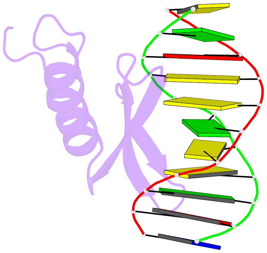
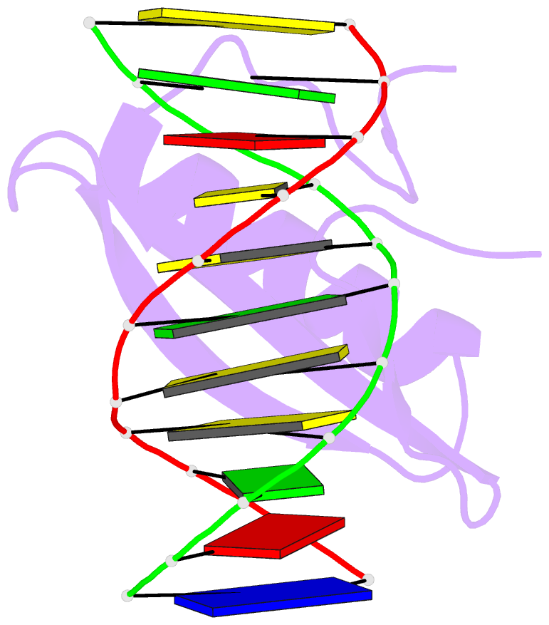
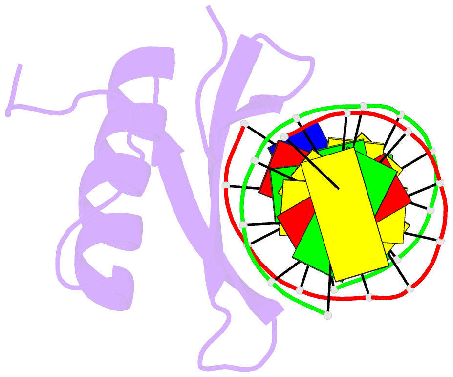
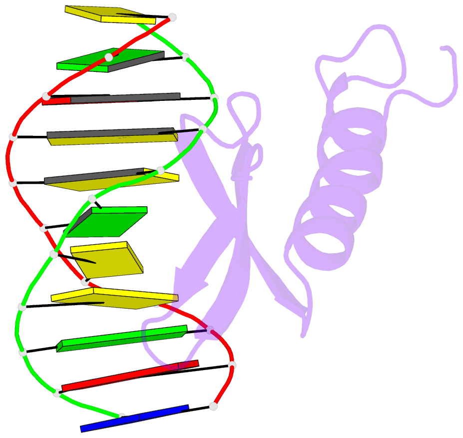
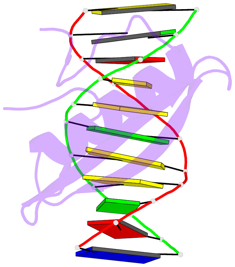
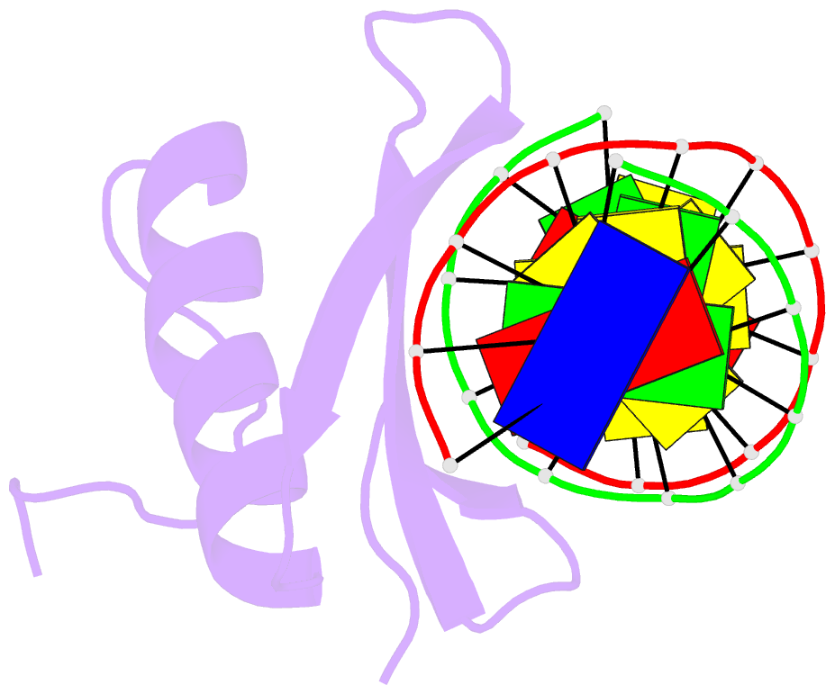
- Solution NMR structure of the complex of gcc-box binding domain of aterf1 and gcc-box DNA, minimized average structure
- Reference
- Allen MD, Yamasaki K, Ohme-Takagi M, Tateno M, Suzuki M (1998): "A novel mode of DNA recognition by a beta-sheet revealed by the solution structure of the GCC-box binding domain in complex with DNA." EMBO J., 17, 5484-5496. doi: 10.1093/emboj/17.18.5484.
- Abstract
- The 3D solution structure of the GCC-box binding domain of a protein from Arabidopsis thaliana in complex with its target DNA fragment has been determined by heteronuclear multidimensional NMR in combination with simulated annealing and restrained molecular dynamic calculation. The domain consists of a three-stranded anti-parallel beta-sheet and an alpha-helix packed approximately parallel to the beta-sheet. Arginine and tryptophan residues in the beta-sheet are identified to contact eight of the nine consecutive base pairs in the major groove, and at the same time bind to the sugar phosphate backbones. The target DNA bends slightly at the central CG step, thereby allowing the DNA to follow the curvature of the beta-sheet.