Summary information and primary citation
- PDB-id
- 1h0m; SNAP-derived features in text and JSON formats;
DNAproDB
- Class
- transcription-DNA
- Method
- X-ray (3.0 Å)
- Summary
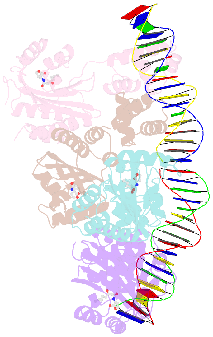
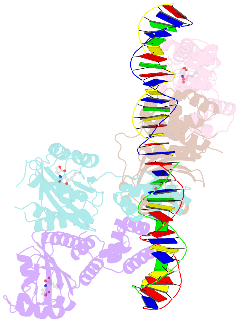
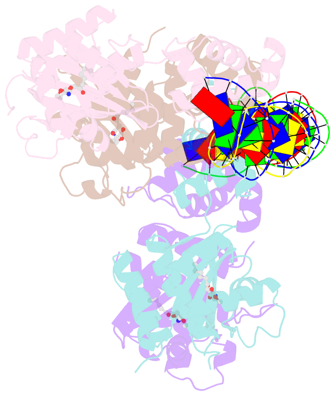
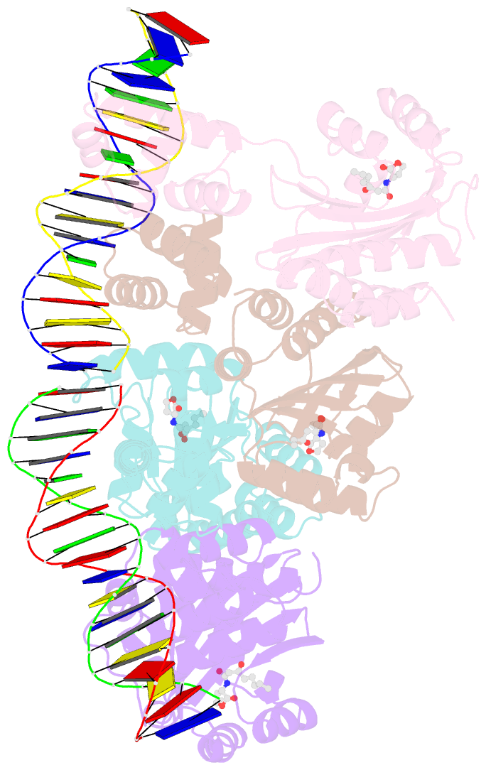
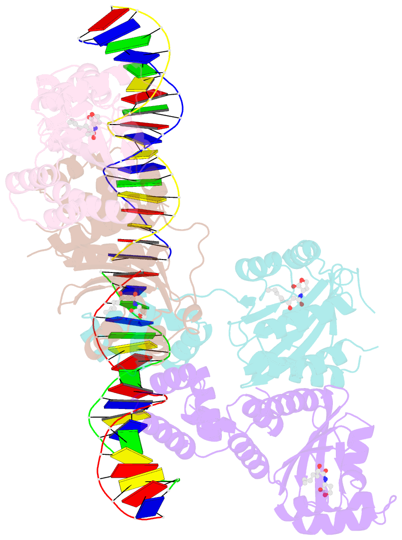
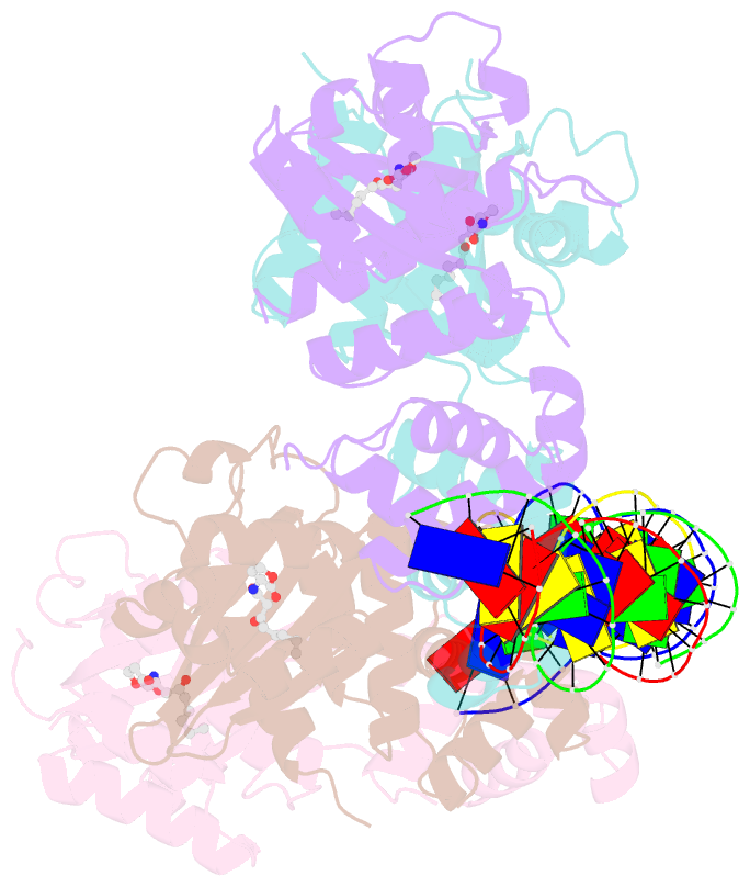
- Three-dimensional structure of the quorum sensing protein trar bound to its autoinducer and to its target DNA
- Reference
- Vannini A, Volpari C, Gargioli C, Muraglia E, Cortese R, De Francesco R, Neddermann P, Di Marco S (2002): "The Crystal Structure of the Quorum Sensing Protein Trar Bound to its Autoinducer and Target DNA." Embo J., 21, 4393. doi: 10.1093/EMBOJ/CDF459.
- Abstract
- The quorum sensing system allows bacteria to sense their cell density and initiate an altered pattern of gene expression after a sufficient quorum of cells has accumulated. In Agrobacterium tumefaciens, quorum sensing controls conjugal transfer of the tumour- inducing plasmid, responsible for plant crown gall disease. The core components of this system are the transcriptional regulator TraR and its inducing ligand N-(3-oxo-octanoyl)-L-homoserine lactone. This complex binds DNA and activates gene expression. We have determined the crystal structure of TraR in complex with its autoinducer and target DNA (PDB code 1h0m). The protein is dimeric, with each monomer composed of an N-terminal domain, which binds the ligand in an enclosed cavity far from the dimerization region, and a C-terminal domain, which binds DNA via a helix-turn-helix motif. The structure reveals an asymmetric homodimer, with one monomer longer than the other. The N-terminal domain resembles GAF/PAS domains, normally fused to catalytic signalling domains. In TraR, the gene fusion is between a GAF/PAS domain and a DNA-binding domain, resulting in a specific transcriptional regulator involved in quorum sensing.