Summary information and primary citation
- PDB-id
- 1h38; SNAP-derived features in text and JSON formats;
DNAproDB
- Class
- transferase
- Method
- X-ray (2.9 Å)
- Summary
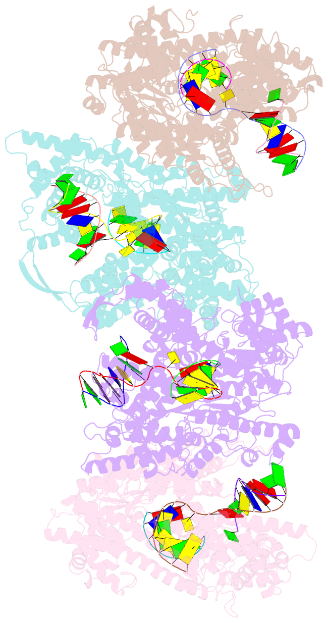
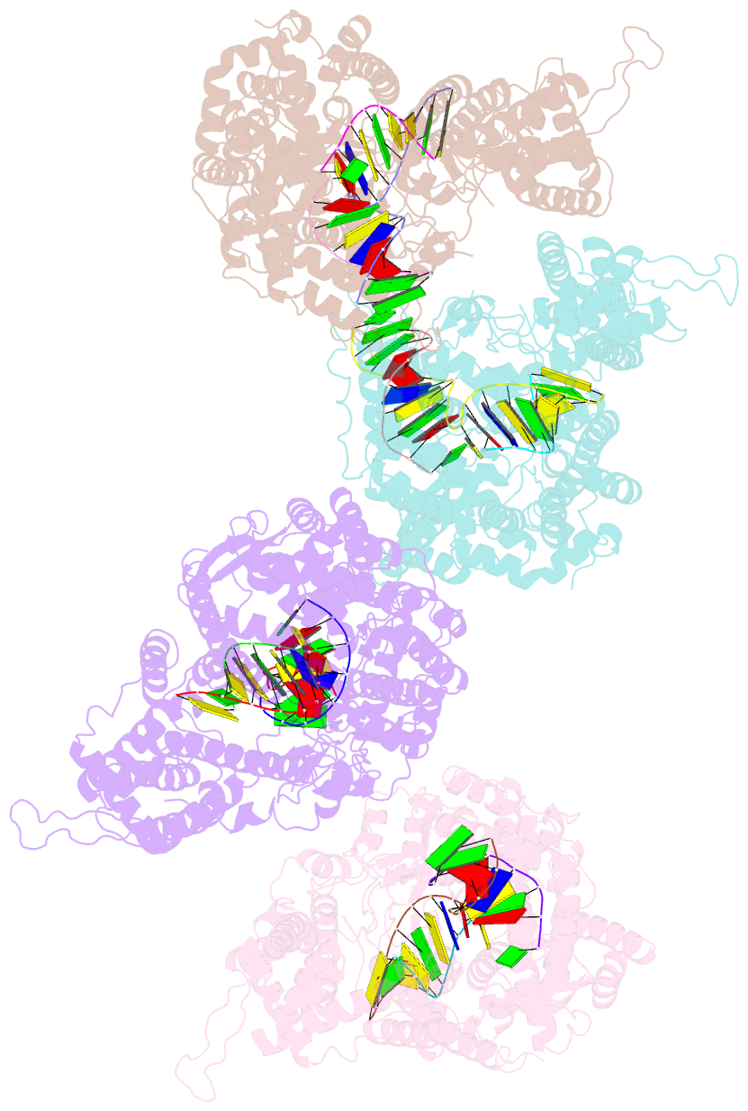
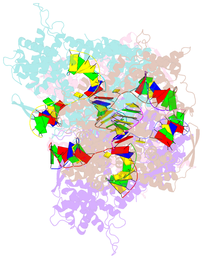
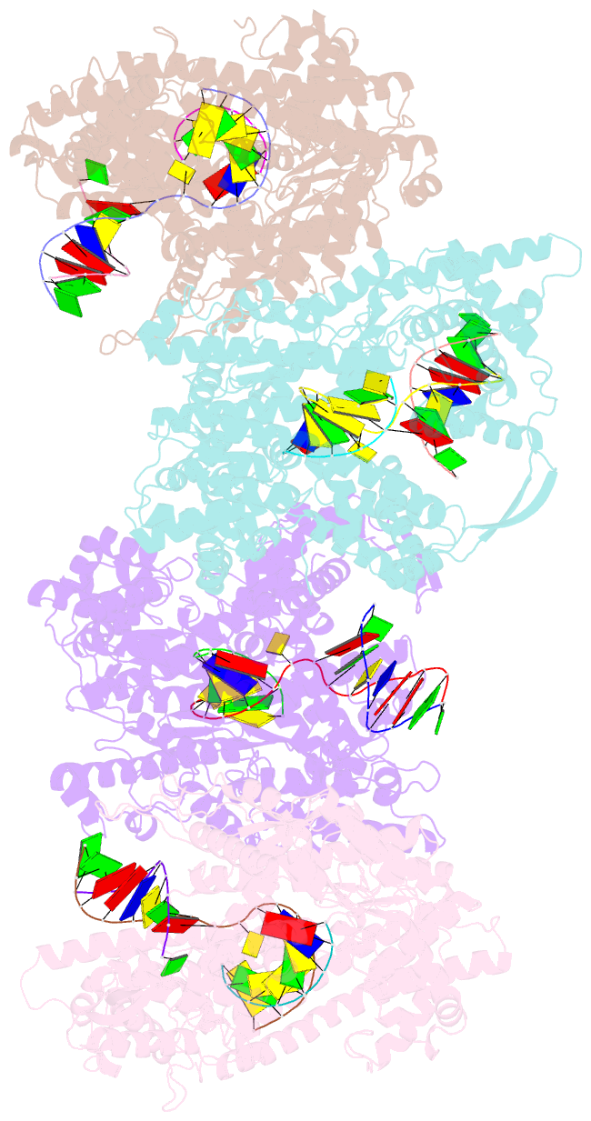
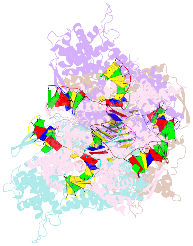
- Structure of a t7 RNA polymerase elongation complex at 2.9a resolution
- Reference
- Tahirov TH, Temiakov D, Anikin M, Patlan V, Mcallister WT, Vassylyev DG, Yokoyama S (2002): "Structure of a T7 RNA Polymerase Elongation Complex at 2.9 A Resolution." Nature, 420, 43. doi: 10.1038/NATURE01129.
- Abstract
- The single-subunit bacteriophage T7 RNA polymerase carries out the transcription cycle in an identical manner to that of bacterial and eukaryotic multisubunit enzymes. Here we report the crystal structure of a T7 RNA polymerase elongation complex, which shows that incorporation of an 8-base-pair RNA-DNA hybrid into the active site of the enzyme induces a marked rearrangement of the amino-terminal domain. This rearrangement involves alternative folding of about 130 residues and a marked reorientation (about 130 degrees rotation) of a stable core subdomain, resulting in a structure that provides elements required for stable transcription elongation. A wide opening on the enzyme surface that is probably an RNA exit pathway is formed, and the RNA-DNA hybrid is completely buried in a newly formed, deep protein cavity. Binding of 10 base pairs of downstream DNA is stabilized mostly by long-distance electrostatic interactions. The structure implies plausible mechanisms for the various phases of the transcription cycle, and reveals important structural similarities with the multisubunit RNA polymerases.