Summary information and primary citation
- PDB-id
- 1h8a; SNAP-derived features in text and JSON formats;
DNAproDB
- Class
- transcription-DNA
- Method
- X-ray (2.23 Å)
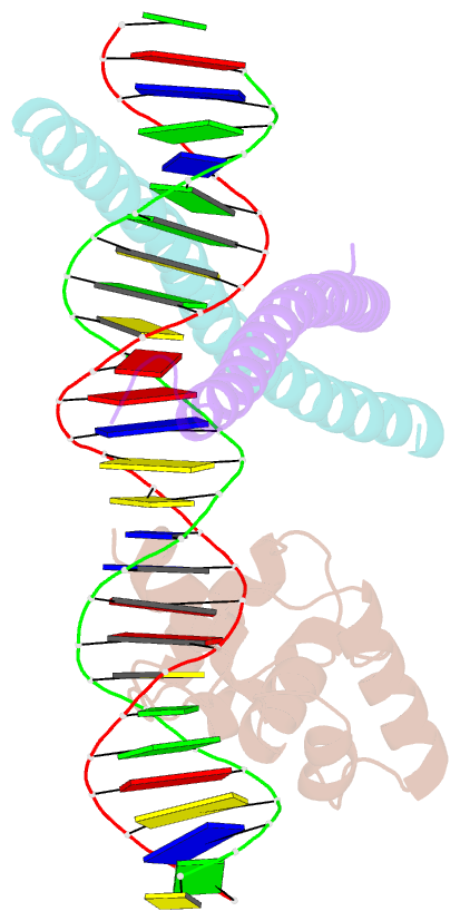
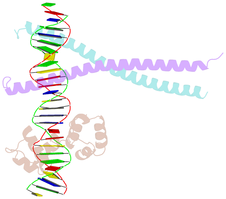
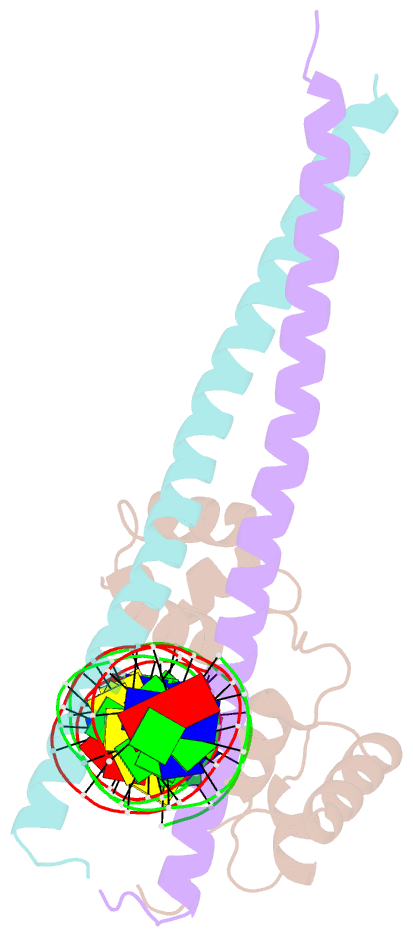
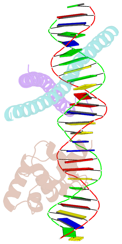
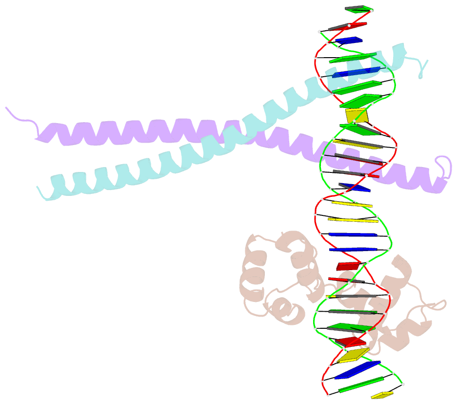
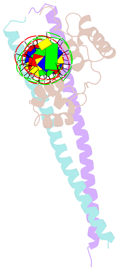
- Summary
- Crystal structure of ternary protein-DNA complex3
- Reference
- Tahirov TH, Sato K, Ichikawa-Iwata E, Sasaki M, Inoue-Bungo T, Shiina M, Kimura K, Takata S, Fujikawa A, Morii H, Kumasaka T, Yamamoto M, Ishii S, Ogata K (2002): "Mechanism of C-Myb-C/Ebpbeta Cooperation from Separated Sites on a Promoter." Cell(Cambridge,Mass.), 108, 57. doi: 10.1016/S0092-8674(01)00636-5.
- Abstract
- c-Myb, but not avian myeloblastosis virus (AMV) v-Myb, cooperates with C/EBP beta to regulate transcription of myeloid-specific genes. To assess the structural basis for that difference, we determined the crystal structures of complexes comprised of the c-Myb or AMV v-Myb DNA-binding domain (DBD), the C/EBP beta DBD, and a promoter DNA fragment. Within the c-Myb complex, a DNA-bound C/EBP beta interacts with R2 of c-Myb bound to a different DNA fragment; point mutations in v-Myb R2 eliminate such interaction within the v-Myb complex. GST pull-down assays, luciferase trans-activation assays, and atomic force microscopy confirmed that the interaction of c-Myb and C/EBP beta observed in crystal mimics their long range interaction on the promoter, which is accompanied by intervening DNA looping.