Summary information and primary citation
- PDB-id
- 1hlo; SNAP-derived features in text and JSON formats;
DNAproDB
- Class
- transcription-DNA
- Method
- X-ray (2.8 Å)
- Summary
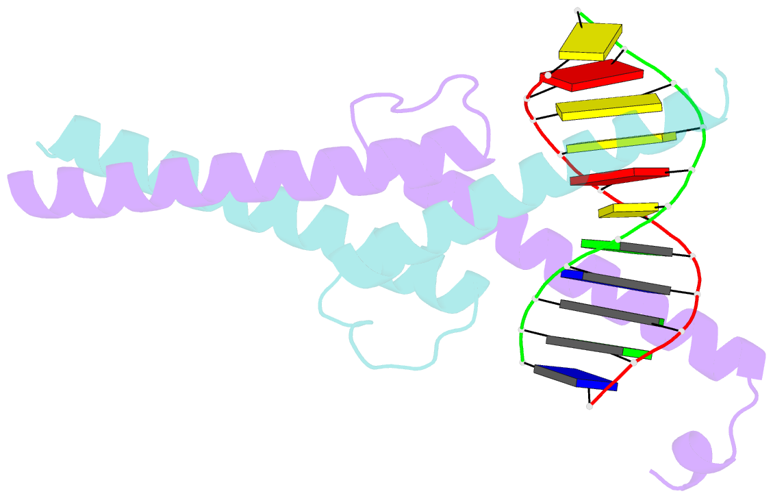
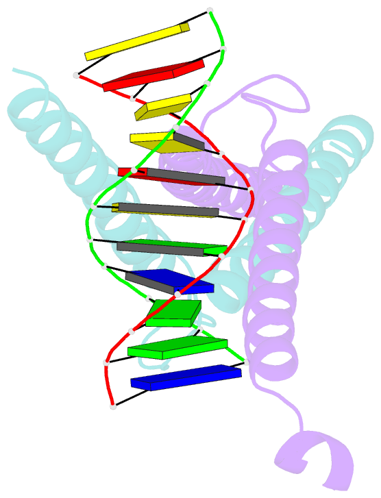
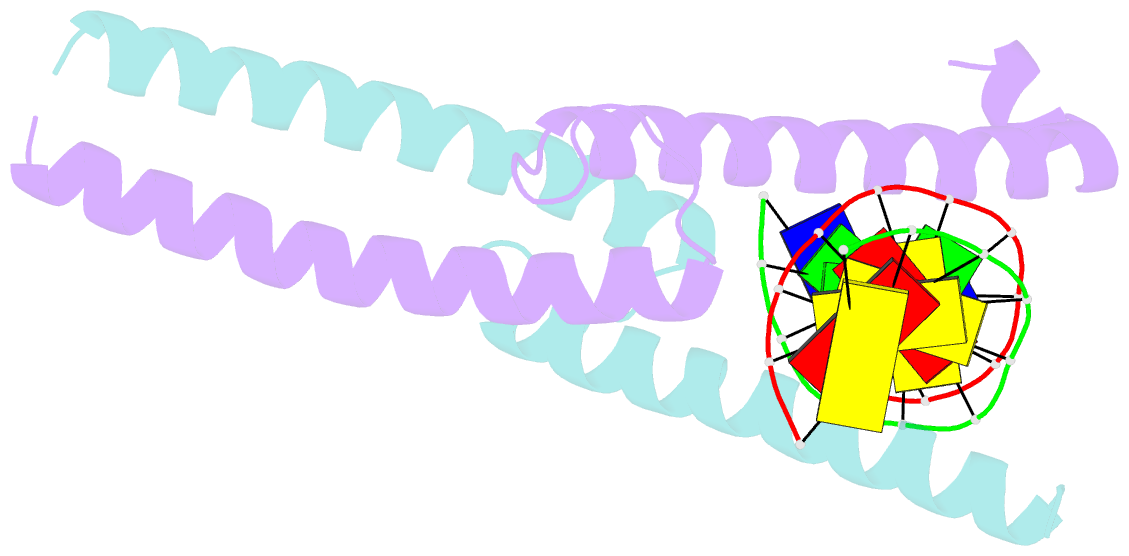
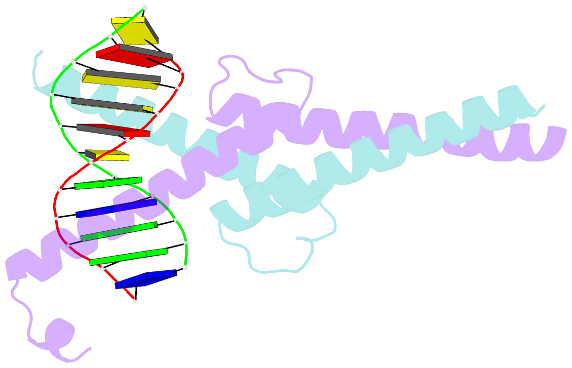
- The crystal structure of an intact human max-DNA complex: new insights into mechanisms of transcriptional control
- Reference
- Brownlie P, Ceska T, Lamers M, Romier C, Stier G, Teo H, Suck D (1997): "The crystal structure of an intact human Max-DNA complex: new insights into mechanisms of transcriptional control." Structure, 5, 509-520. doi: 10.1016/S0969-2126(97)00207-4.
- Abstract
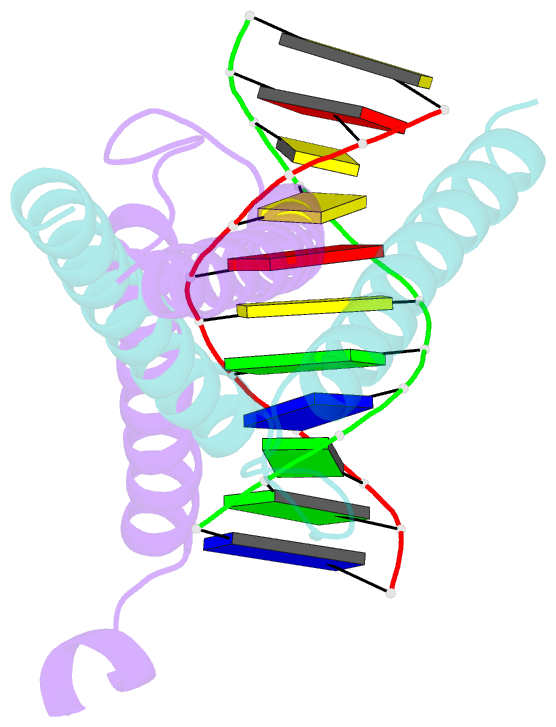
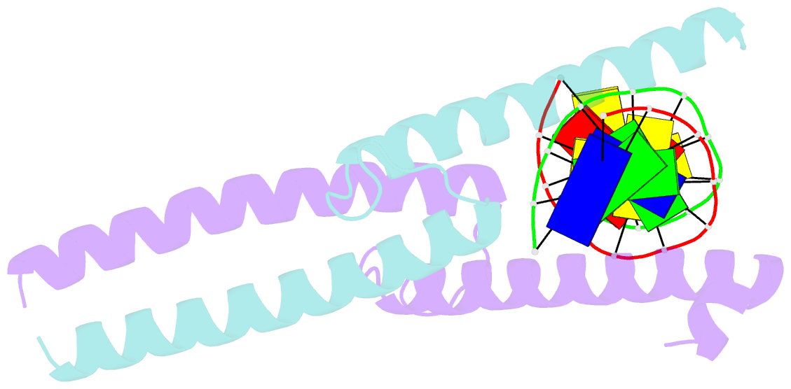
- Background: Max belongs to the basic helix-loop-helix leucine zipper (bHLHZ) family of transcription factors. Max is able to form homodimers and heterodimers with other members of this family, which include Mad, Mxi1 and Myc; Myc is an oncoprotein implicated in cell proliferation, differentiation and apoptosis. The homodimers and heterodimers compete for a common DNA target site (the E box) and rearrangement amongst these dimer forms provides a complex system of transcriptional regulation. Max is also regulated by phosphorylation at a site preceding the basic region. We report here the first crystal structure of an intact bHLHZ protein bound to its target site.
Results: The X-ray crystal structure of the intact human Max protein homodimer in complex with a 13-mer DNA duplex was determined to 2.8 A resolution and refined to an R factor of 0.213. The C-terminal domains in both chains of the Max dimer are disordered. In contrast to the DNA observed in complex with other bHLH and bHLHZ proteins, the DNA in the Max complex is bent by about 25 degrees, directed towards the protein. Intimate contacts with interdigitating sidechains give rise to the formation of tetramers in the crystal.
Conclusions: The structure confirms the importance of the HLH and leucine zipper motifs in dimerization as well as the mode of E box recognition which was previously analyzed by X-ray crystallography of shortened constructs. The disorder observed in the C-terminal domain suggests that contacts with additional protein components of the transcription machinery are necessary for ordering the secondary structure. The tetramers seen in the crystal are consistent with the tendency of Max and other bHLHZ and HLH proteins to form higher order oligomers in solution and may play a role in DNA looping. The location of the two phosphorylation sites at Ser1 and Ser10 (the latter is the N-cap of the basic helix) suggests how phosphorylation could disrupt DNA binding.