Summary information and primary citation
- PDB-id
- 1i3j; SNAP-derived features in text and JSON formats;
DNAproDB
- Class
- hydrolase-DNA
- Method
- X-ray (2.2 Å)
- Summary
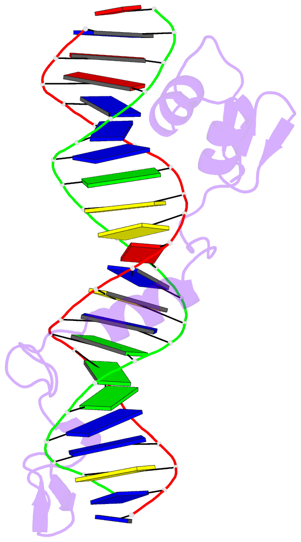
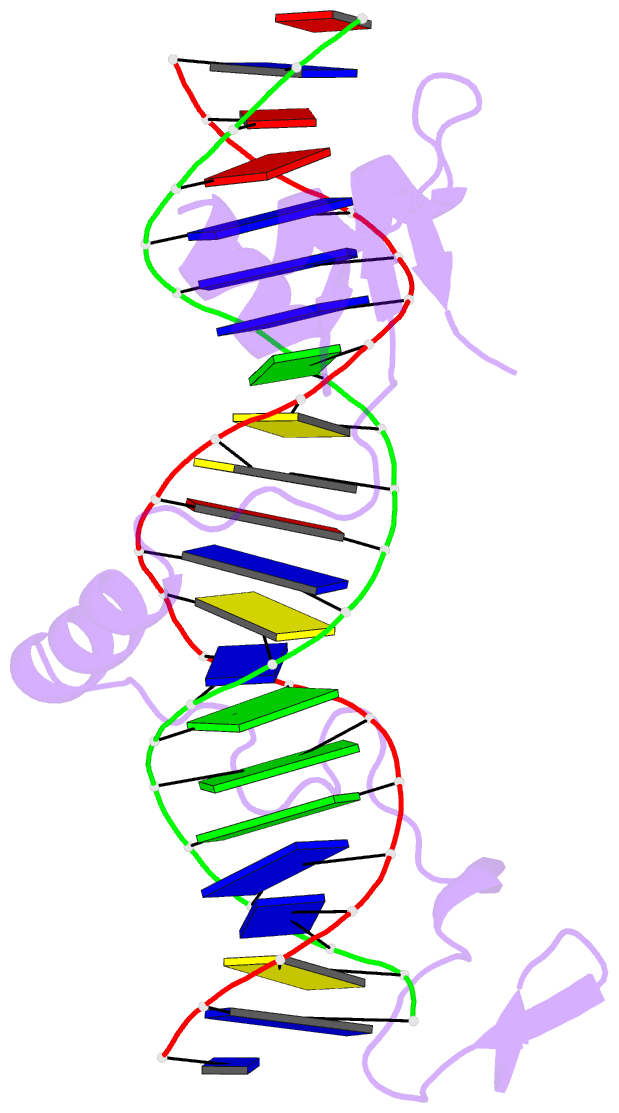
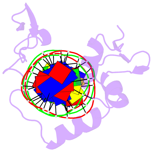
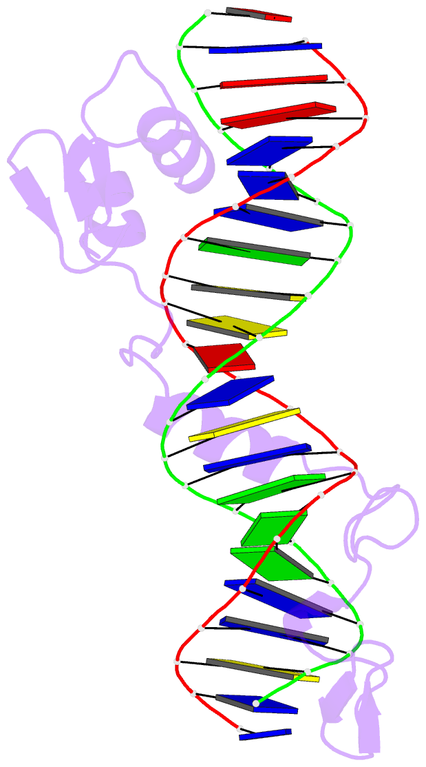
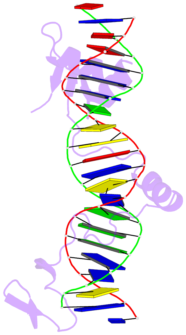
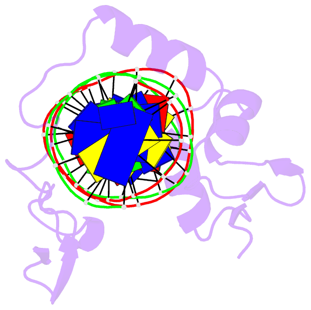
- Crystal structure of the DNA-binding domain of intron endonuclease i-tevi with its substrate
- Reference
- Van Roey P, Waddling CA, Fox KM, Belfort M, Derbyshire V (2001): "Intertwined structure of the DNA-binding domain of intron endonuclease I-TevI with its substrate." EMBO J., 20, 3631-3637. doi: 10.1093/emboj/20.14.3631.
- Abstract
- I-TevI is a site-specific, sequence-tolerant intron endonuclease. The crystal structure of the DNA-binding domain of I-TevI complexed with the 20 bp primary binding region of its DNA target reveals an unusually extended structure composed of three subdomains: a Zn finger, an elongated segment containing a minor groove-binding alpha-helix, and a helix-turn-helix. The protein wraps around the DNA, mostly following the minor groove, contacting the phosphate backbone along the full length of the duplex. Surprisingly, while the minor groove-binding helix and the helix-turn- helix subdomain make hydrophobic contacts, the few base-specific hydrogen bonds occur in segments that lack secondary structure and flank the intron insertion site. The multiple base-specific interactions over a long segment of the substrate are consistent with the observed high site specificity in spite of sequence tolerance, while the modular composition of the domain is pertinent to the evolution of homing endonucleases.