Summary information and primary citation
- PDB-id
- 1i3w; SNAP-derived features in text and JSON formats;
DNAproDB
- Class
- DNA-antibiotic
- Method
- X-ray (1.7 Å)
- Summary
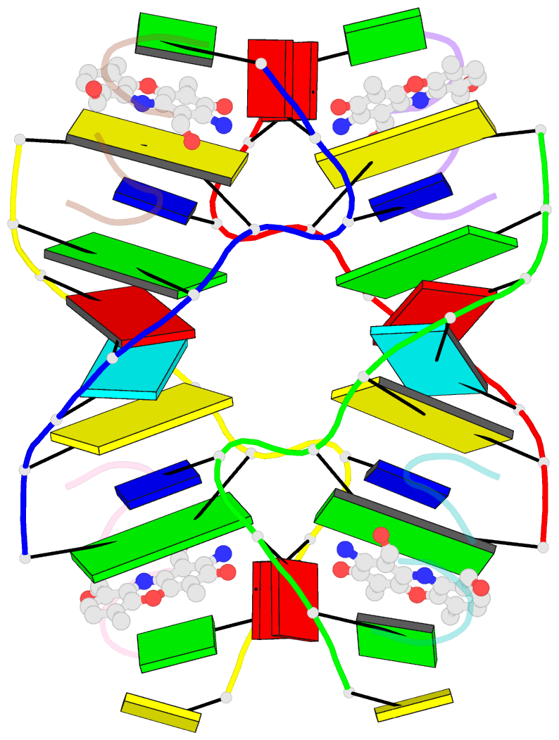
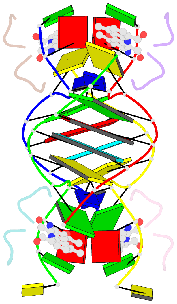
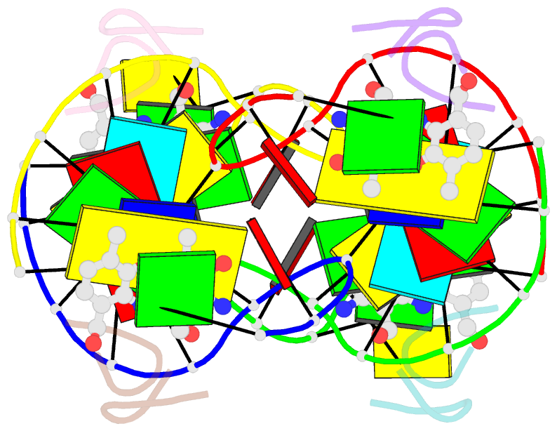
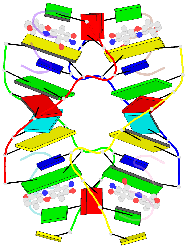
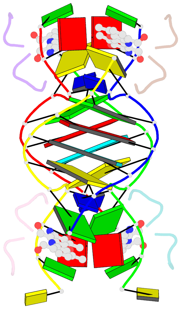
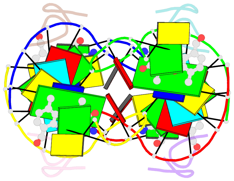
- Actinomycin d binding to cgatcgatcg
- Reference
- Robinson H, Gao YG, Yang X, Sanishvili R, Joachimiak A, Wang AH (2001): "Crystallographic Analysis of a Novel Complex of Actinomycin D Bound to the DNA Decamer Cgatcgatcg." Biochemistry, 40, 5587. doi: 10.1021/BI002859Z.
- Abstract
- The potent anticancer drug actinomycin D (ActD) acts by binding to DNA, thereby interfering with replication and transcription. ActD inhibits RNA polymerase far more specifically than DNA polymerase. Such discrimination is not easily understood by the conventional DNA binding mode of ActD. We have solved and refined at 1.7 A resolution the crystal structure of ActD complexed to CGATCGATCG, which contains no canonical GpC binding sequence. The crystal data are space group P4(3)2(1)2, a = b = 47.01 A, and c = 160.37 A. The structure was solved by the multiple wavelength anomalous diffraction method using a 5-bromo-U DNA. The asymmetric unit of the unit cell contains two independent dimers of a novel slipped duplex complex consisting of two decamer DNA strands bound with two ActD drug molecules. (The DNA in one dimer is numbered C1 to G10 in one strand and C11 to G20 in the complementary strand and in the second dimer, C101 to G110 and C111 to G120, respectively.) The structure reveals a highly unusual ActD binding mode in which the DNA adopts a slipped duplex with the A3-T4/A13-T14 dinucleotides looped out. ActD intercalates between G2-C11* (C11* being from a symmetry-related molecule) and C5-G20 base pairs. Two such slipped duplex-ActD complexes bound to each other by mutually intercalating their T4/T14 bases into the helix cavities (located between C5-G20 and G6-C19 base pairs) of neighboring complexes, forming a dimer of drug-DNA complexes. The binding site mimics the drug binding at the elongation point during transcription. Modeling studies show that the ActD-DNA complex fits snugly in the active site cavity in RNA polymerase but not in DNA polymerase. This may explain the strong preference of ActD inhibition toward transcription.