Summary information and primary citation
- PDB-id
- 1ig7; SNAP-derived features in text and JSON formats;
DNAproDB
- Class
- transcription-DNA
- Method
- X-ray (2.2 Å)
- Summary
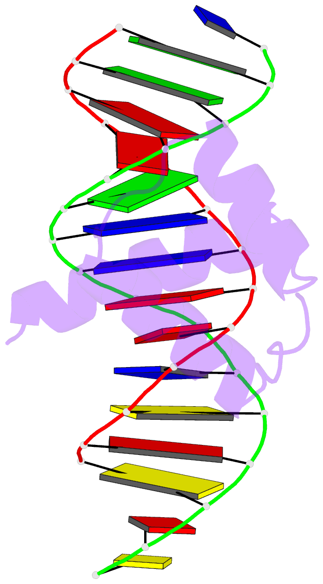
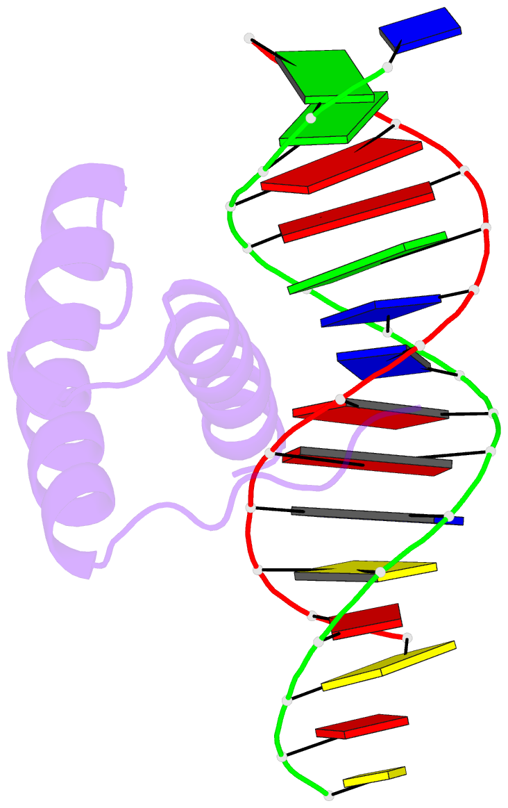
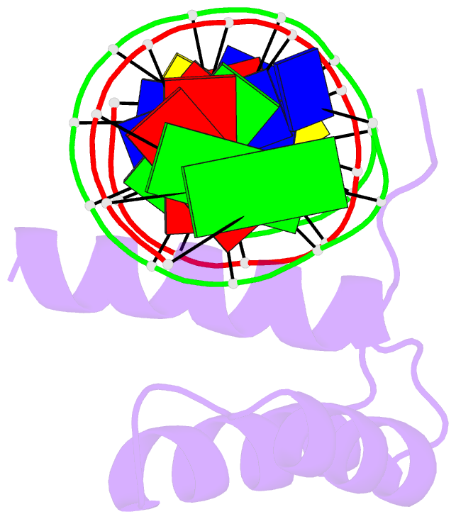
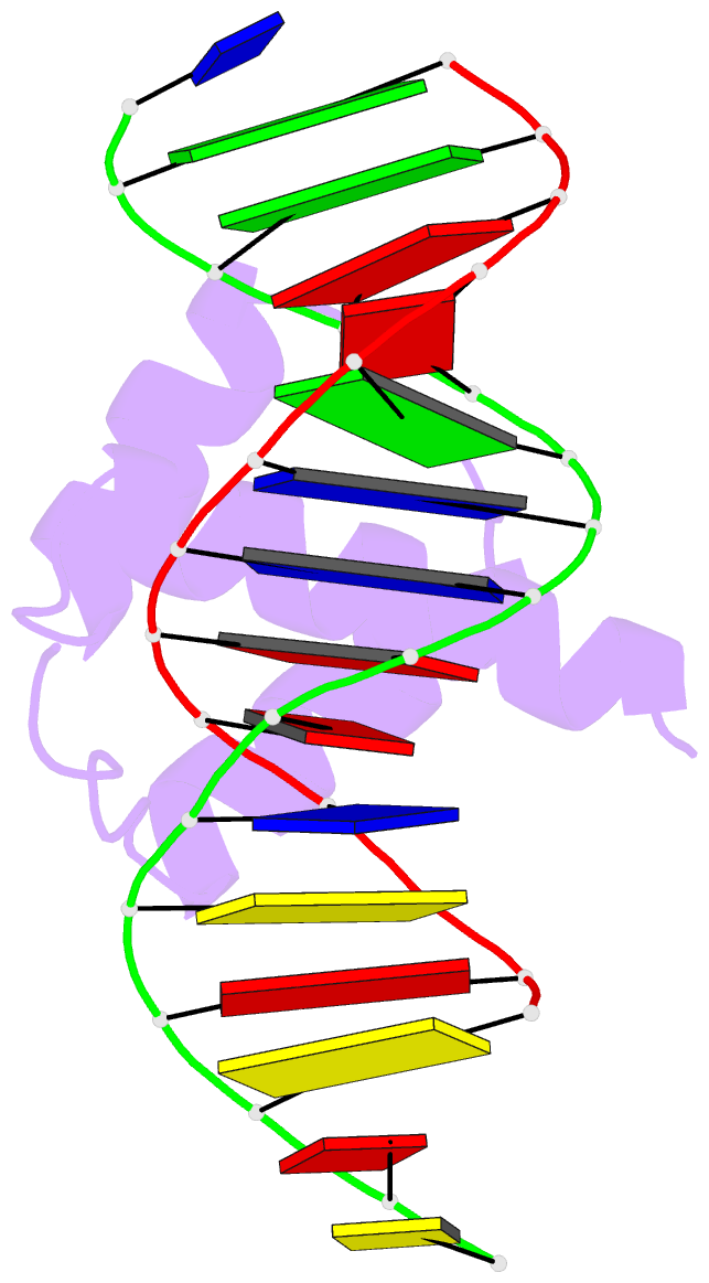
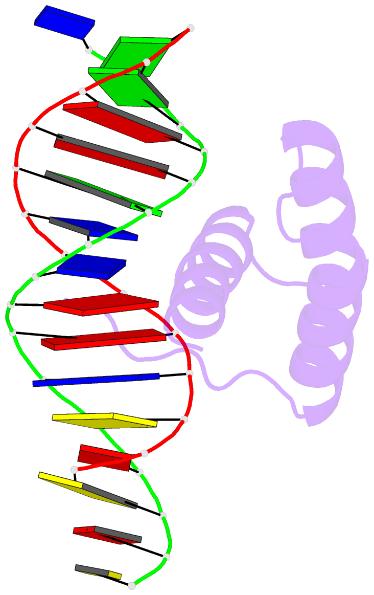
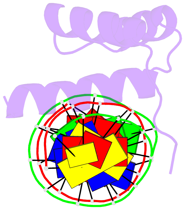
- Msx-1 homeodomain-DNA complex structure
- Reference
- Hovde S, Abate-Shen C, Geiger JH (2001): "Crystal structure of the Msx-1 homeodomain/DNA complex." Biochemistry, 40, 12013-12021. doi: 10.1021/bi0108148.
- Abstract
- The Msx-1 homeodomain protein plays a crucial role in craniofacial, limb, and nervous system development. Homeodomain DNA-binding domains are comprised of 60 amino acids that show a high degree of evolutionary conservation. We have determined the structure of the Msx-1 homeodomain complexed to DNA at 2.2 A resolution. The structure has an unusually well-ordered N-terminal arm with a unique trajectory across the minor groove of the DNA. DNA specificity conferred by bases flanking the core TAAT sequence is explained by well ordered water-mediated interactions at Q50. Most interactions seen at the TAAT sequence are typical of the interactions seen in other homeodomain structures. Comparison of the Msx-1-HD structure to all other high resolution HD-DNA complex structures indicate a remarkably well-conserved sphere of hydration between the DNA and protein in these complexes.