Summary information and primary citation
- PDB-id
- 1jmc; SNAP-derived features in text and JSON formats;
DNAproDB
- Class
- replication-DNA
- Method
- X-ray (2.4 Å)
- Summary
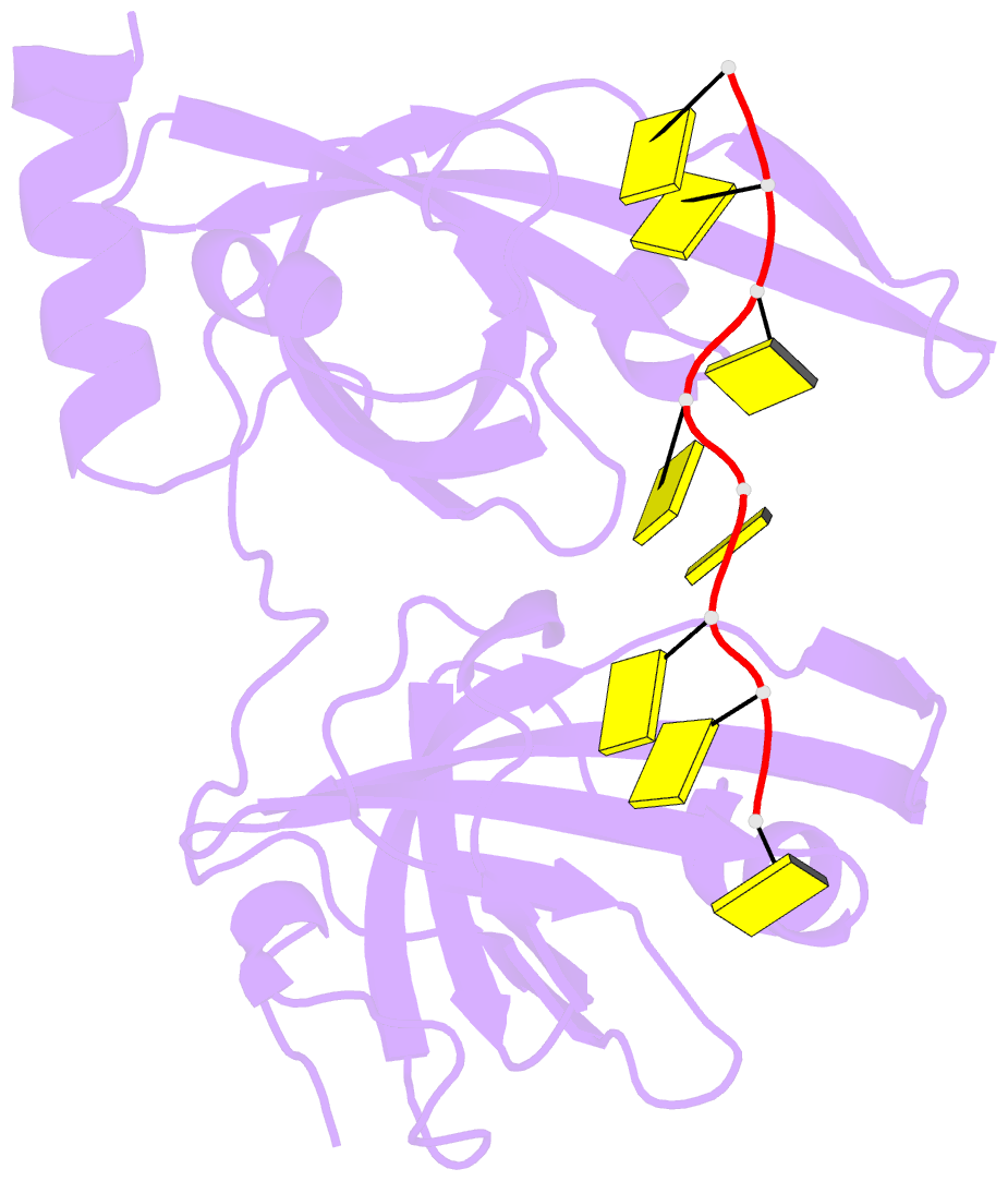
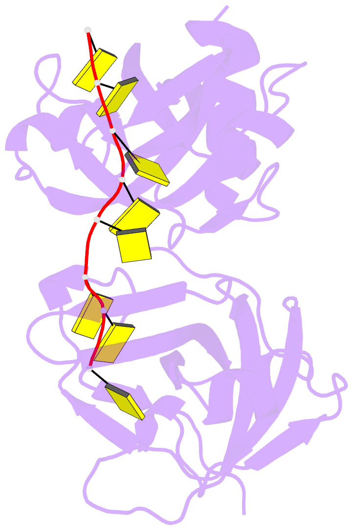
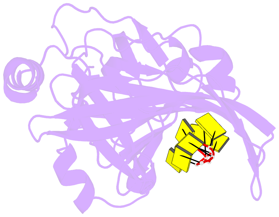
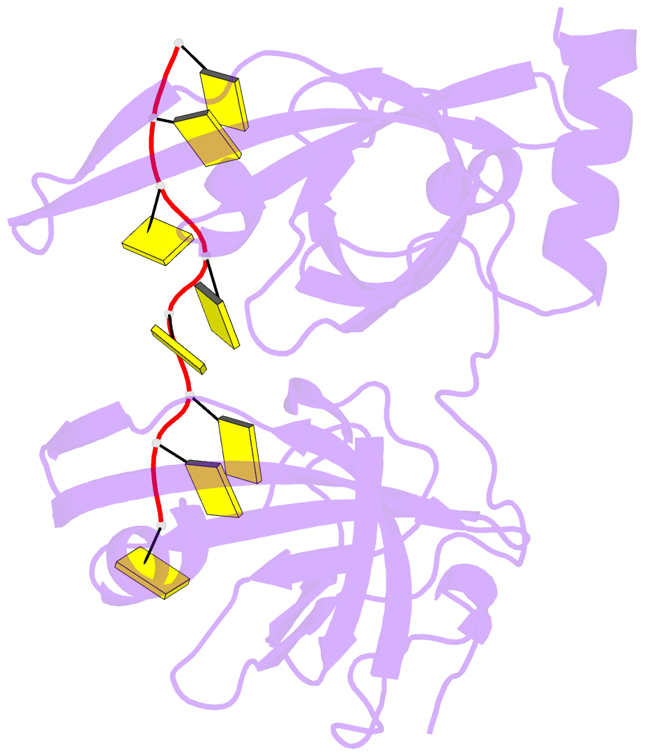
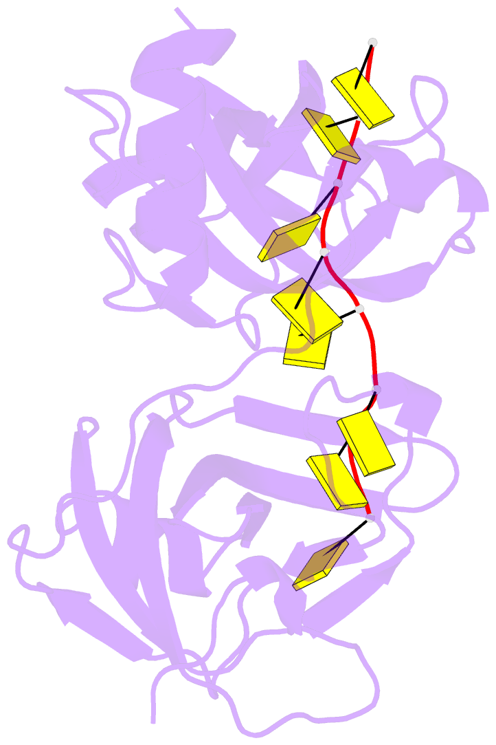
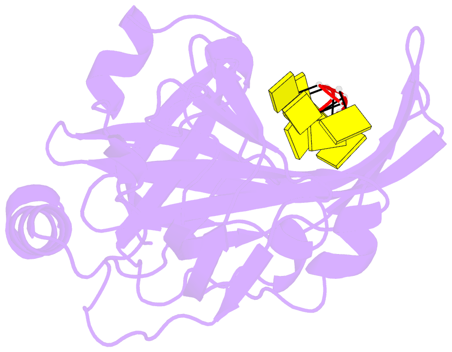
- Single stranded DNA-binding domain of human replication protein a bound to single stranded DNA, rpa70 subunit, residues 183-420
- Reference
- Bochkarev A, Pfuetzner RA, Edwards AM, Frappier L (1997): "Structure of the single-stranded-DNA-binding domain of replication protein A bound to DNA." Nature, 385, 176-181. doi: 10.1038/385176a0.
- Abstract
- The single-stranded-DNA-binding proteins (SSBs) are essential for DNA function in prokaryotic and eukaryotic cells, mitochondria, phages and viruses. The structures of four SSBs have been solved, but the molecular details of the interaction of SSBs with DNA remain speculative. We report here the crystal structure at 2.4 A resolution of the single-stranded-DNA-binding domain of human replication protein A (RPA) bound to DNA. Replication protein A is a heterotrimeric SSB that is highly conserved in eukaryotes. The largest subunit, RPA70, binds to single-stranded (ss)DNA and mediates interactions with many cellular and viral proteins. The DNA-binding domain, which lies in the middle of RPA70, comprises two structurally homologous subdomains oriented in tandem. The ssDNA lies in a channel that extends from one subdomain to the other. The structure of each RPA70 subdomain is similar to those of the bacteriophage SSBs, indicating that the mechanism of ssDNA-binding is conserved.