Summary information and primary citation
- PDB-id
- 1jwl; SNAP-derived features in text and JSON formats;
DNAproDB
- Class
- transcription-DNA
- Method
- X-ray (4.0 Å)
- Summary
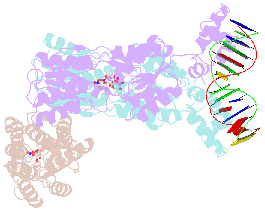
- Structure of the dimeric lac repressor-operator o1-onpf complex
- Reference
- Bell CE, Lewis M (2001): "Crystallographic analysis of Lac repressor bound to natural operator O1." J.Mol.Biol., 312, 921-926. doi: 10.1006/jmbi.2001.5024.
- Abstract
- Previous structures of Lac repressor bound to DNA used a fully symmetric "ideal" operator sequence that is missing the central G-C base-pair present in the three natural operator sequences. Here we have determined the X-ray crystal structure of a dimeric Lac repressor bound to a 22 base-pair DNA with the natural operator O1 sequence and the anti-inducer ONPF, at 4.0 A resolution. The natural operator is bent in the same way as the symmetric sequence, due to the binding of the hinge helices of the repressor to the minor groove at the central GCGG sequence of O1. Comparison of the structures of the repressor bound to the natural and symmetric operators shows very similar overall structures, with only slight rearrangements of the headpiece domains of the repressor. Analysis of crystals with iodinated DNA shows that the operator is uniquely positioned and allows for the sequence registration of the DNA relative to the repressor to be determined. The kink in the operator is centered between the left half-site and the central G-C base-pair of O1. Our results are most consistent with a previously proposed model in which, relative to the complex with the symmetric operator, the repressor accommodates binding to the natural operator sequence by shifting the position of the right headpiece by one base-pair step towards the center of O1.


