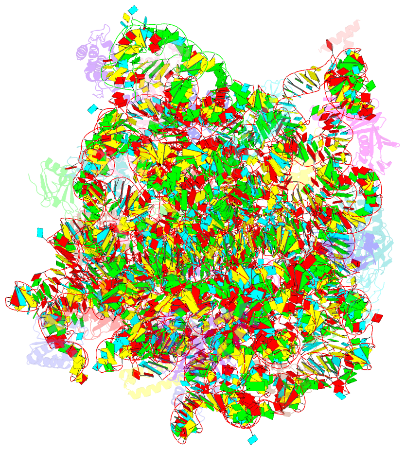
Summary information and primary citation
- PDB-id
- 1kc8; SNAP-derived features in text and JSON formats;
DNAproDB
- Class
- ribosome
- Method
- X-ray (3.01 Å)
- Summary
- Co-crystal structure of blasticidin s bound to the 50s ribosomal subunit
- Reference
- Hansen J, Moore PB, Steitz TA (2003): "Structures of Five Antibiotics Bound at the Peptidyl Transferase Center of
the Large Ribosomal Subunit." J.Mol.Biol., 330, 1061-1075. doi: 10.1016/S0022-2836(03)00668-5.
- Abstract
- Structures of anisomycin, chloramphenicol, sparsomycin, blasticidin S, and virginiamycin M bound to the large ribosomal subunit of Haloarcula marismortui have been determined at 3.0A resolution. Most of these antibiotics bind to sites that overlap those of either peptidyl-tRNA or aminoacyl-tRNA, consistent with their functioning as competitive inhibitors of peptide bond formation. Two hydrophobic crevices, one at the peptidyl transferase center and the other at the entrance to the peptide exit tunnel play roles in binding these antibiotics. Midway between these crevices, nucleotide A2103 of H.marismortui (2062 Escherichia coli) varies in its conformation and thereby contacts antibiotics bound at either crevice. The aromatic ring of anisomycin binds to the active-site hydrophobic crevice, as does the aromatic ring of puromycin, while the aromatic ring of chloramphenicol binds to the exit tunnel hydrophobic crevice. Sparsomycin contacts primarily a P-site bound substrate, but also extends into the active-site hydrophobic crevice. Virginiamycin M occupies portions of both the A and P-site, and induces a conformational change in the ribosome. Blasticidin S base-pairs with the P-loop and thereby mimics C74 and C75 of a P-site bound tRNA.


