Summary information and primary citation
- PDB-id
- 1kfv; SNAP-derived features in text and JSON formats;
DNAproDB
- Class
- hydrolase-DNA
- Method
- X-ray (2.55 Å)
- Summary
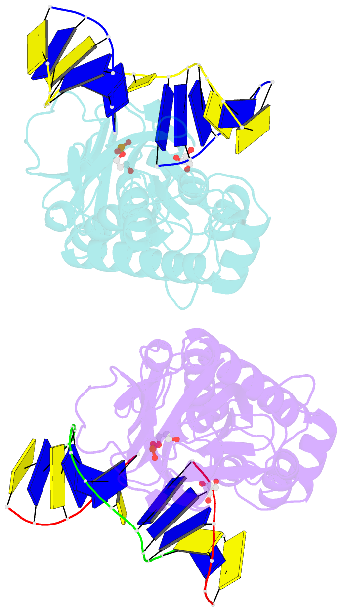
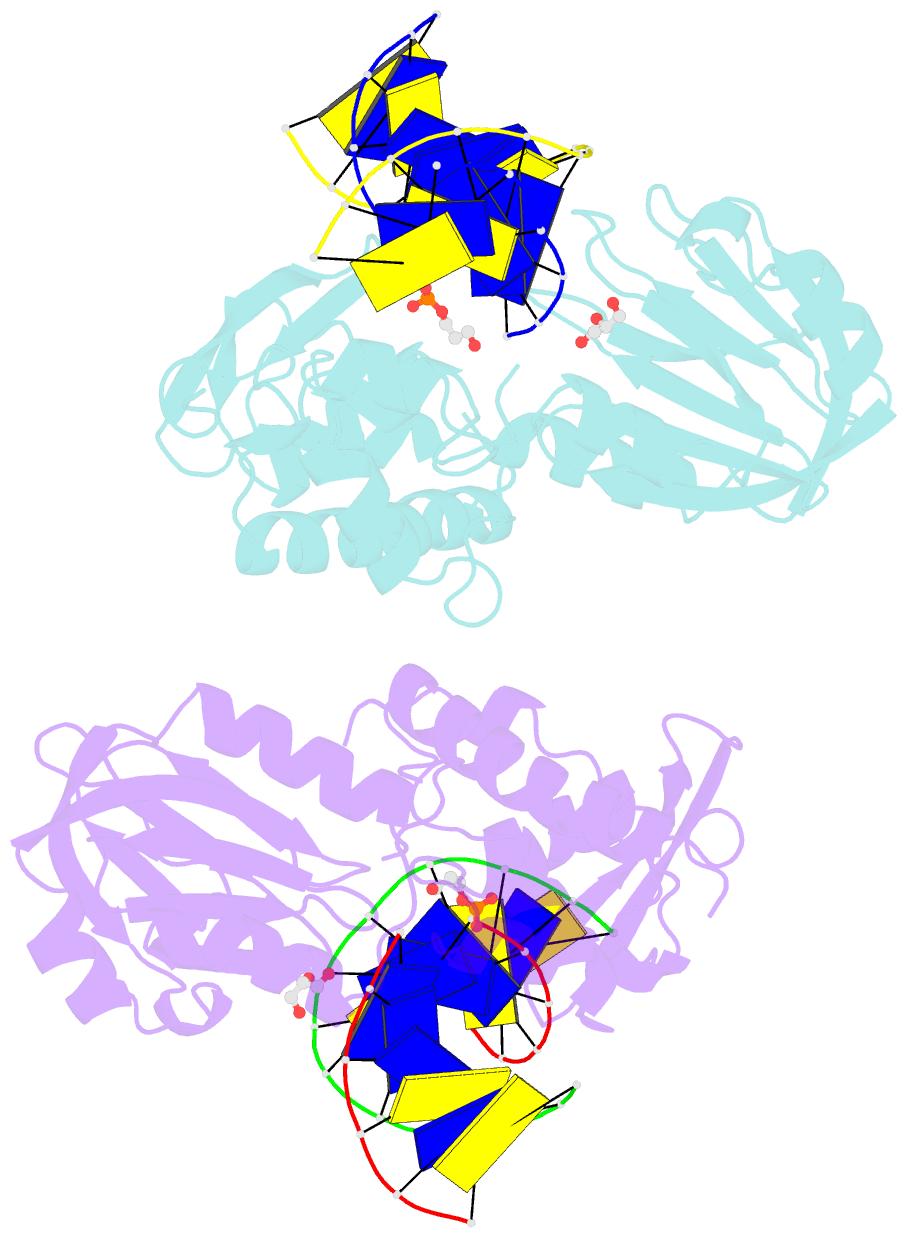
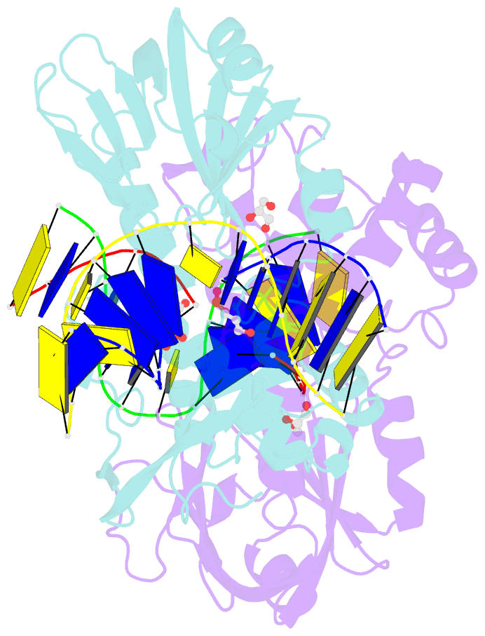
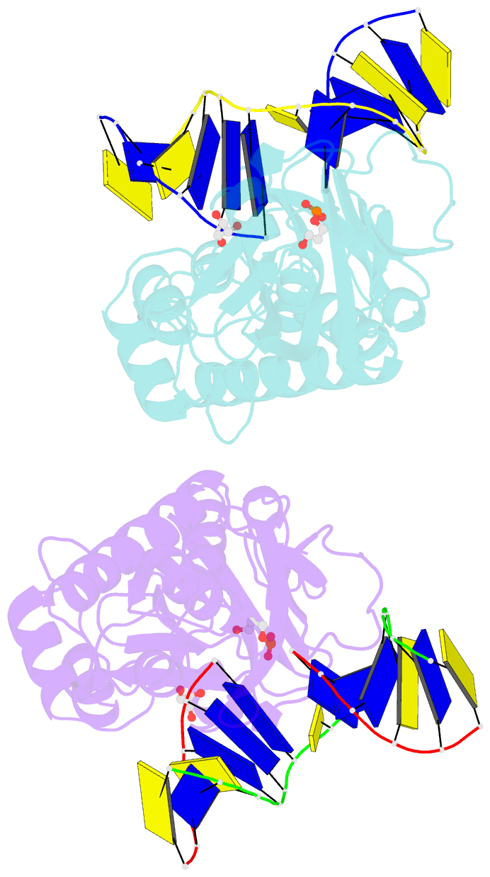
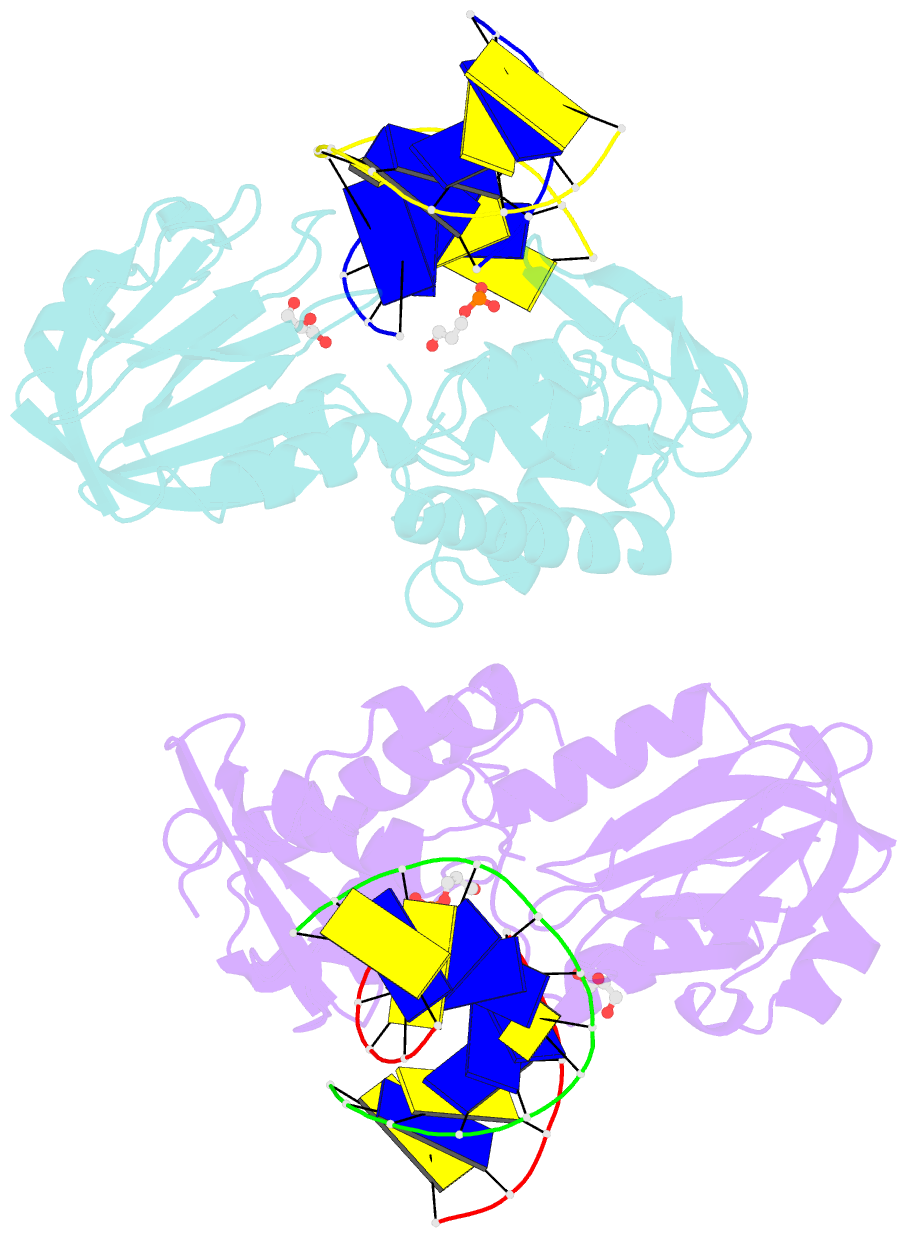
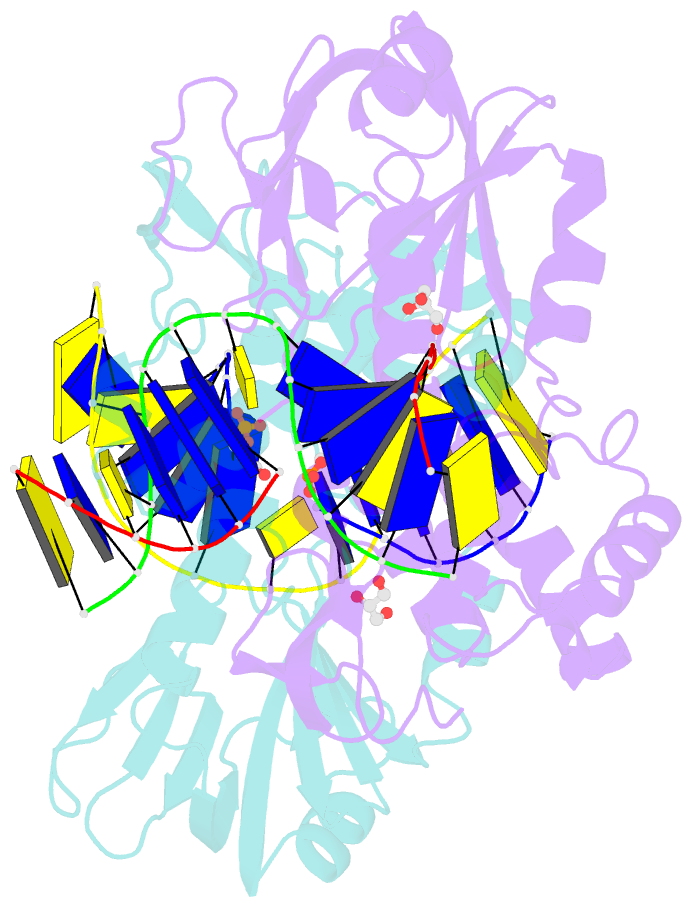
- Crystal structure of lactococcus lactis formamido-pyrimidine DNA glycosylase (alias fpg or mutm) non covalently bound to an ap site containing DNA.
- Reference
- Serre L, Pereira de Jesus K, Boiteux S, Zelwer C, Castaing B (2002): "Crystal structure of the Lactococcus lactis formamidopyrimidine-DNA glycosylase bound to an abasic site analogue-containing DNA." EMBO J., 21, 2854-2865. doi: 10.1093/emboj/cdf304.
- Abstract
- The formamidopyrimidine-DNA glycosylase (Fpg, MutM) is a bifunctional base excision repair enzyme (DNA glycosylase/AP lyase) that removes a wide range of oxidized purines, such as 8-oxoguanine and imidazole ring-opened purines, from oxidatively damaged DNA. The structure of a non-covalent complex between the Lactoccocus lactis Fpg and a 1,3-propanediol (Pr) abasic site analogue-containing DNA has been solved. Through an asymmetric interaction along the damaged strand and the intercalation of the triad (M75/R109/F111), Fpg pushes out the Pr site from the DNA double helix, recognizing the cytosine opposite the lesion and inducing a 60 degrees bend of the DNA. The specific recognition of this cytosine provides some structural basis for understanding the divergence between Fpg and its structural homologue endo nuclease VIII towards their substrate specificities. In addition, the modelling of the 8-oxoguanine residue allows us to define an enzyme pocket that may accommodate the extrahelical oxidized base.