Summary information and primary citation
- PDB-id
- 1lcd; SNAP-derived features in text and JSON formats;
DNAproDB
- Class
- gene regulation-DNA
- Method
- NMR
- Summary
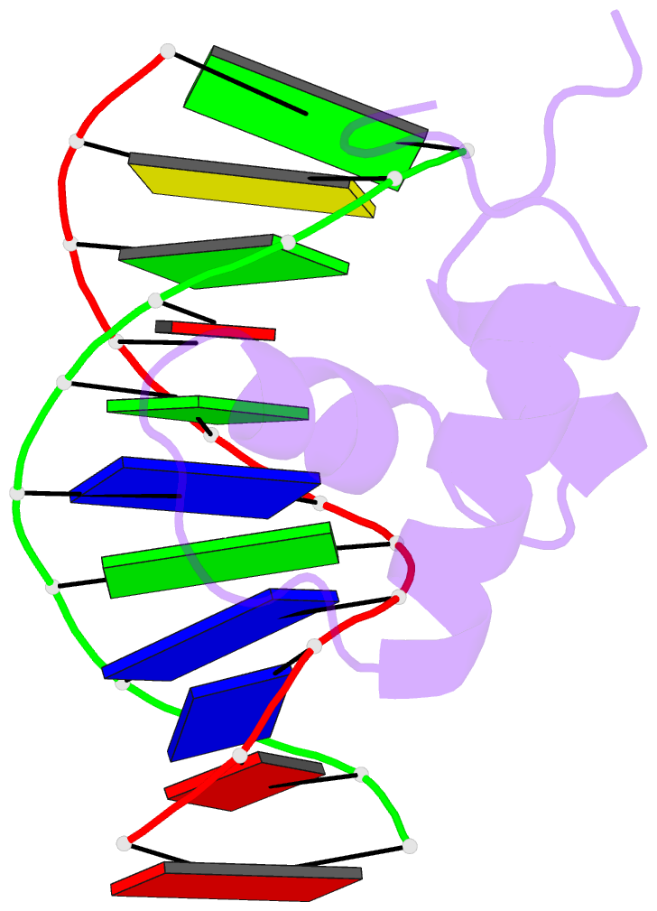
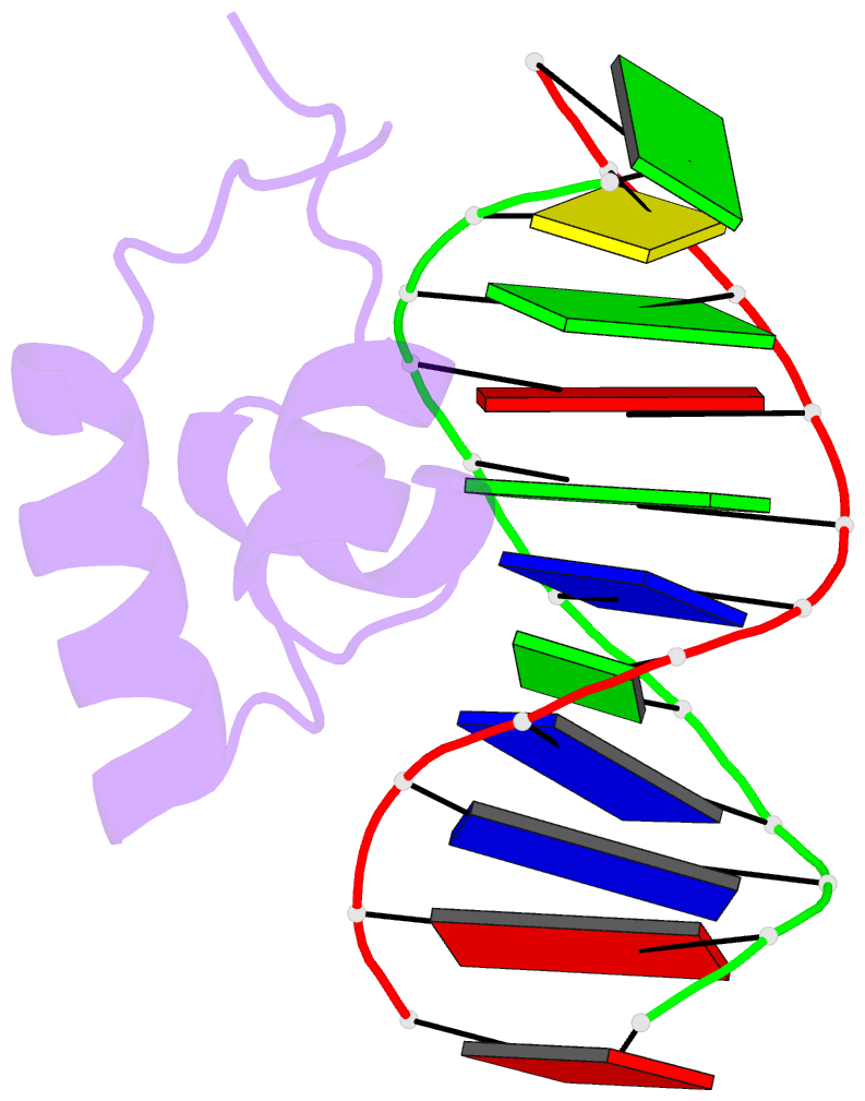
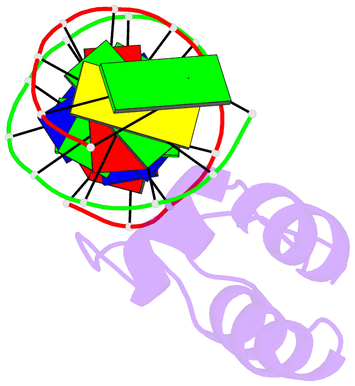
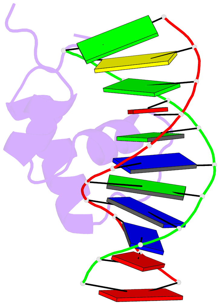
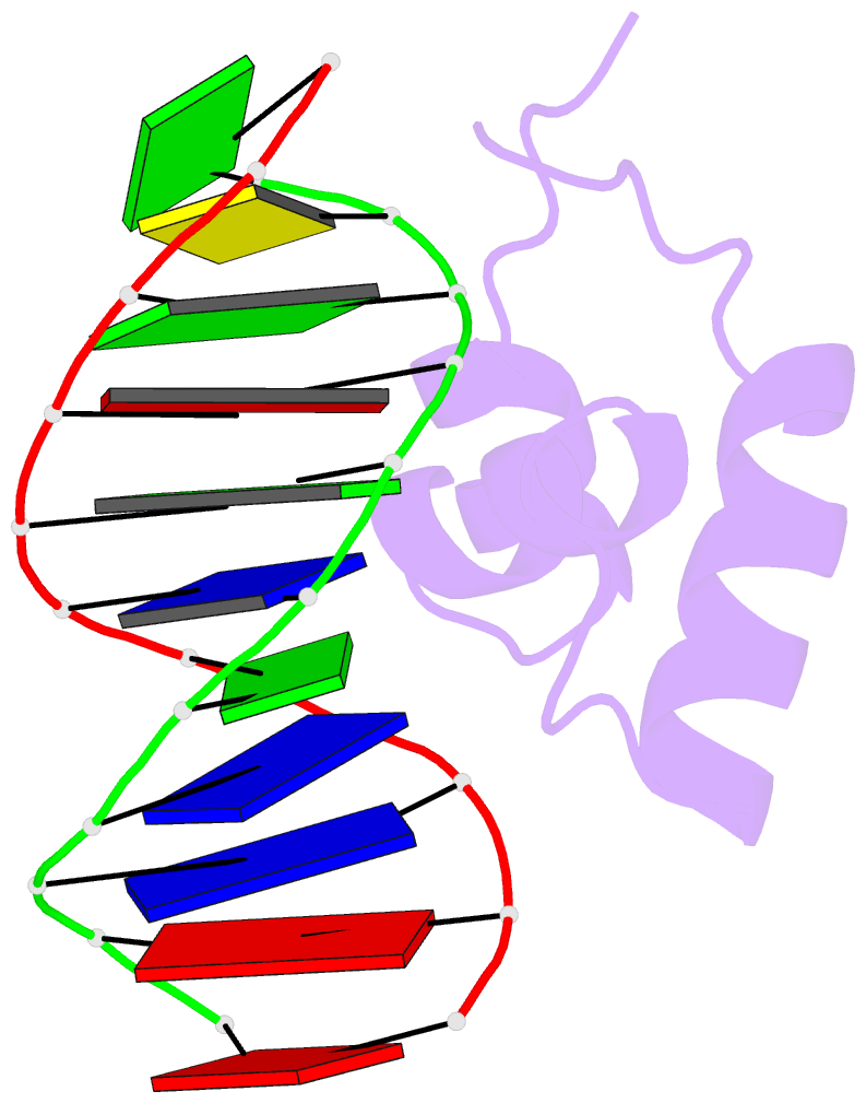
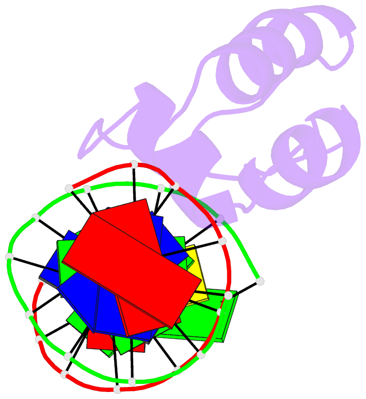
- Structure of the complex of lac repressor headpiece and an 11 base-pair half-operator determined by nuclear magnetic resonance spectroscopy and restrained molecular dynamics
- Reference
- Chuprina VP, Rullmann JA, Lamerichs RM, van Boom JH, Boelens R, Kaptein R (1993): "Structure of the complex of lac repressor headpiece and an 11 base-pair half-operator determined by nuclear magnetic resonance spectroscopy and restrained molecular dynamics." J.Mol.Biol., 234, 446-462. doi: 10.1006/jmbi.1993.1598.
- Abstract
- The structure of the complex of lac repressor headpiece and an 11 base-pair lac half-operator has been determined by NMR spectroscopy and restrained Molecular Dynamics calculations. In total 508 distances were derived from two-dimensional nuclear Overhauser enhancement measurements, 260 of which are within the headpiece, 212 within the operator and 36 between operator and headpiece. An equilibrium restrained Molecular Dynamics calculation of the complex in aqueous solution, spanning 85 picoseconds, has been used to analyze the structure. Configuration sampling by an annealing procedure has been undertaken as well in order to estimate the precision of the structure determination. Our data confirm the results of previous two-dimensional NMR studies that the orientation of the recognition helix of lac repressor in the major groove of DNA with respect to the operator dyad axis is opposite to the orientation found in complexes of other DNA binding proteins of the helix-turn-helix class. We find a number of tight contacts between the protein and the operator that are in agreement with the available genetic and biochemical data. The anchoring of lac headpiece on the operator is similar to that of other repressors. Other features are unique for lac headpiece: relative few direct hydrogen bonds between side-chains and bases; extensive apolar contacts; many direct and water-bridged contacts to phosphates from residues in or close to the recognition helix. Overall, an interconnected set of interactions is observed, involving base-specific contacts, phosphate contacts, intra-protein and water-bridged hydrogen bonds. Several of these interactions appear to be dynamic, i.e. fluctuating in time, rather than static.