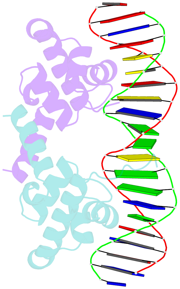
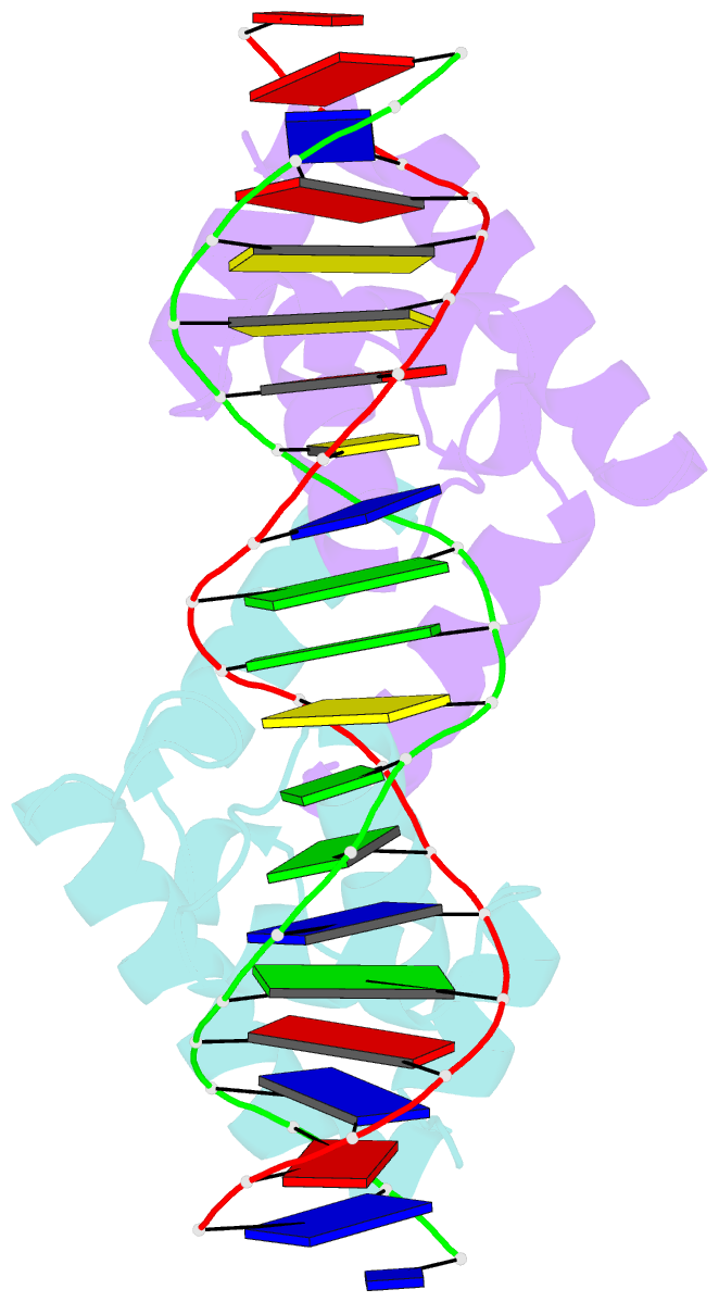
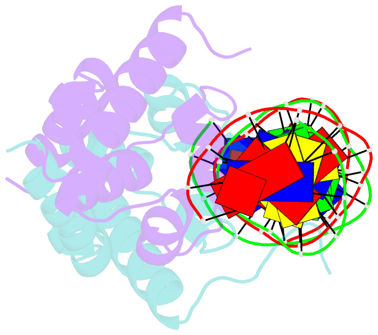
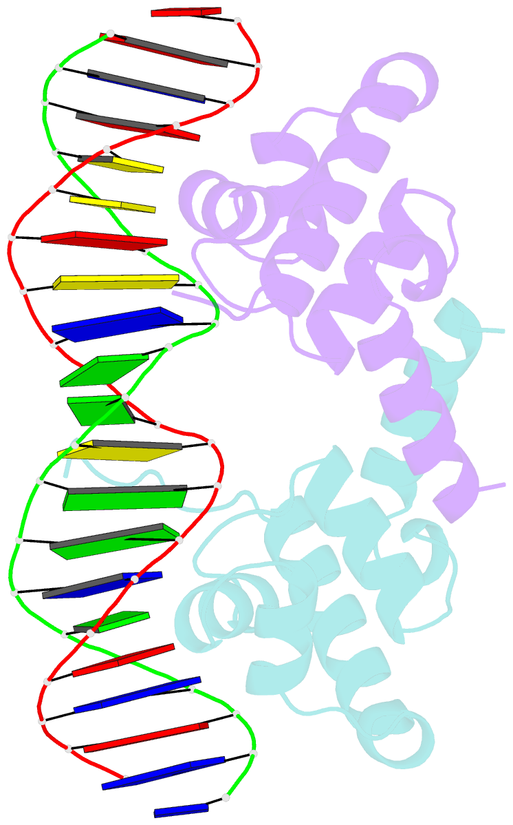
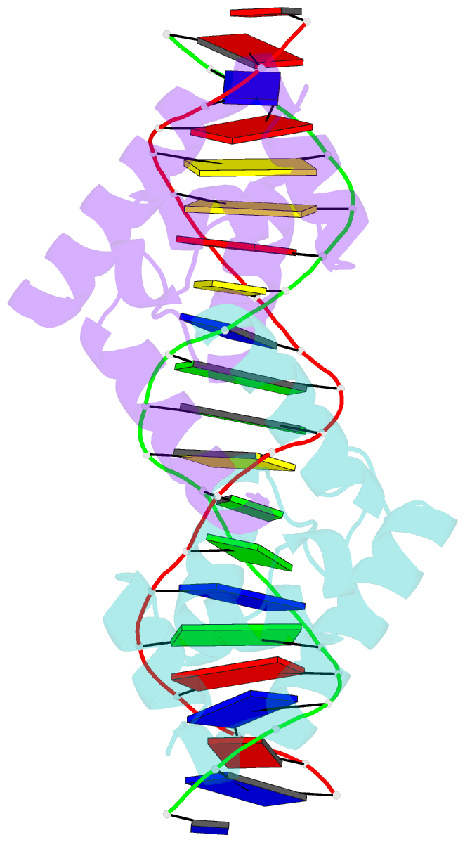
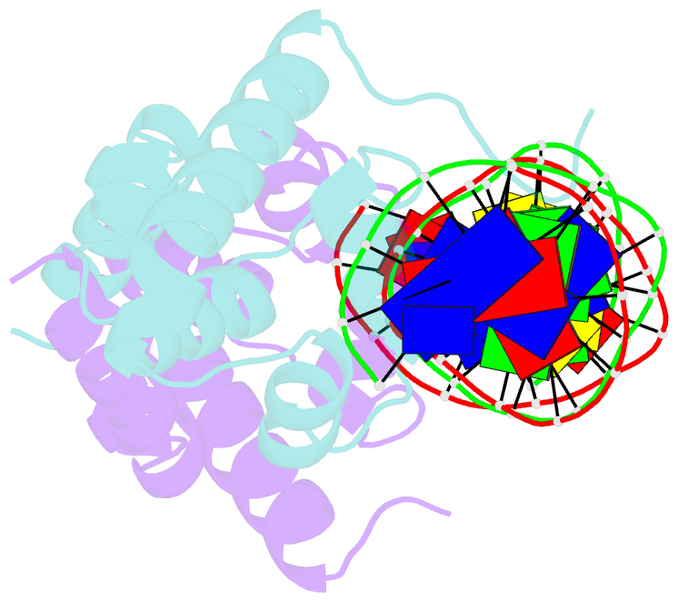
Summary information and primary citation
- PDB-id
- 1lmb; SNAP-derived features in text and JSON formats;
DNAproDB
- Class
- transcription-DNA
- Method
- X-ray (1.8 Å)
- Summary
- Refined 1.8 angstrom crystal structure of the lambda repressor-operator complex
- Reference
- Beamer LJ, Pabo CO (1992): "Refined 1.8 A crystal structure of the lambda repressor-operator complex." J.Mol.Biol., 227, 177-196. doi: 10.1016/0022-2836(92)90690-L.
- Abstract
- The crystal structure of the lambda repressor-operator complex has been refined to an R-factor of 18.9% at 1.8 A resolution. This refinement, using data collected at low temperature, has revealed the structure of the N-terminal arm and shows that the interactions of repressor with the two halves of the pseudo-symmetric operator site are significantly different. The two halves of the complex are most similar near the outer edge of the operator site (in a region where the lambda and 434 repressors make similar contacts), but they become increasingly different toward the center of the operator. There are striking differences near the center of the site where it appears that the arm makes significant contacts to only one half of the DNA site. This suggested a new way of aligning the operator sites in phage lambda. The high resolution structure confirms many of the previously noted features of the complex, but also reveals a number of new protein-DNA contacts. It also gives a better view of the extensive H-bonding networks that couple contacts made by different residues and different regions of the protein, and reveals important new details about the helix-turn-helix (HTH) region, and the positions of many water molecules in the complex.