Summary information and primary citation
- PDB-id
- 1m1k; SNAP-derived features in text and JSON formats;
DNAproDB
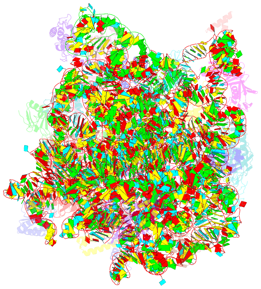
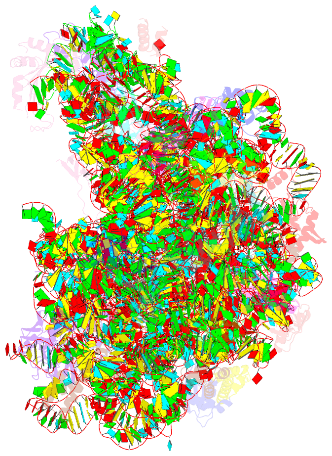
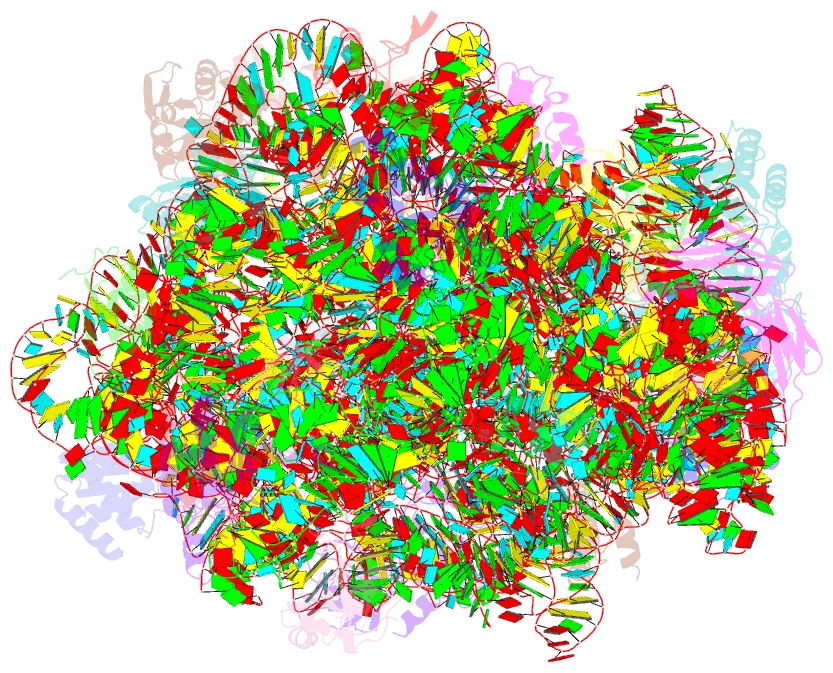
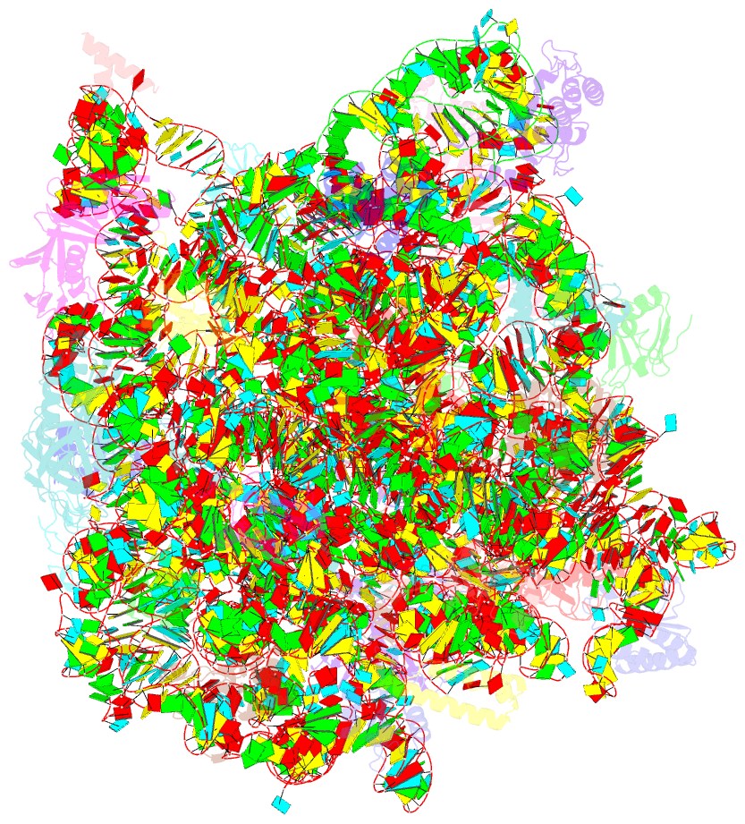
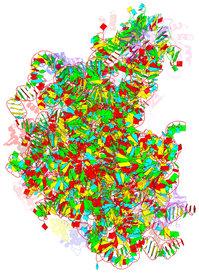
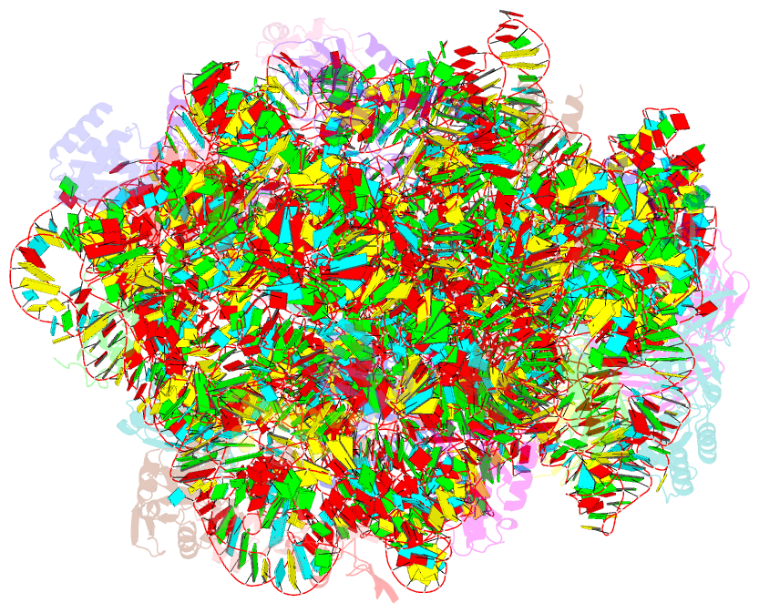
- Class
- ribosome
- Method
- X-ray (3.2 Å)
- Summary
- Co-crystal structure of azithromycin bound to the 50s ribosomal subunit of haloarcula marismortui
- Reference
- Hansen JL, Ippolito JA, Ban N, Nissen P, Moore PB, Steitz TA (2002): "The structures of four macrolide antibiotics bound to the large ribosomal subunit." Mol.Cell, 10, 117-128. doi: 10.1016/S1097-2765(02)00570-1.
- Abstract
- Crystal structures of the Haloarcula marismortui large ribosomal subunit complexed with the 16-membered macrolide antibiotics carbomycin A, spiramycin, and tylosin and a 15-membered macrolide, azithromycin, show that they bind in the polypeptide exit tunnel adjacent to the peptidyl transferase center. Their location suggests that they inhibit protein synthesis by blocking the egress of nascent polypeptides. The saccharide branch attached to C5 of the lactone rings extends toward the peptidyl transferase center, and the isobutyrate extension of the carbomycin A disaccharide overlaps the A-site. Unexpectedly, a reversible covalent bond forms between the ethylaldehyde substituent at the C6 position of the 16-membered macrolides and the N6 of A2103 (A2062, E. coli). Mutations in 23S rRNA that result in clinical resistance render the binding site less complementary to macrolides.