Summary information and primary citation
- PDB-id
- 1mzp; SNAP-derived features in text and JSON formats;
DNAproDB
- Class
- ribosome
- Method
- X-ray (2.65 Å)
- Summary
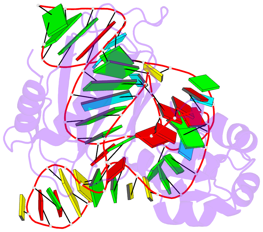
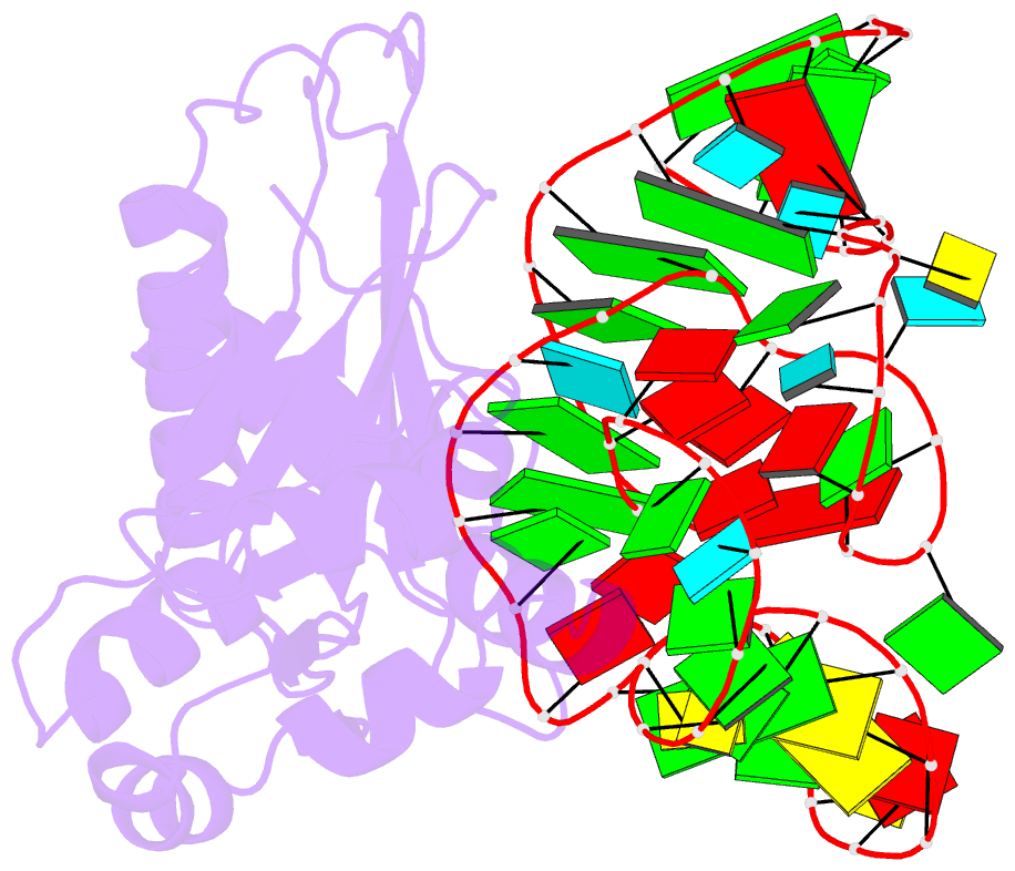
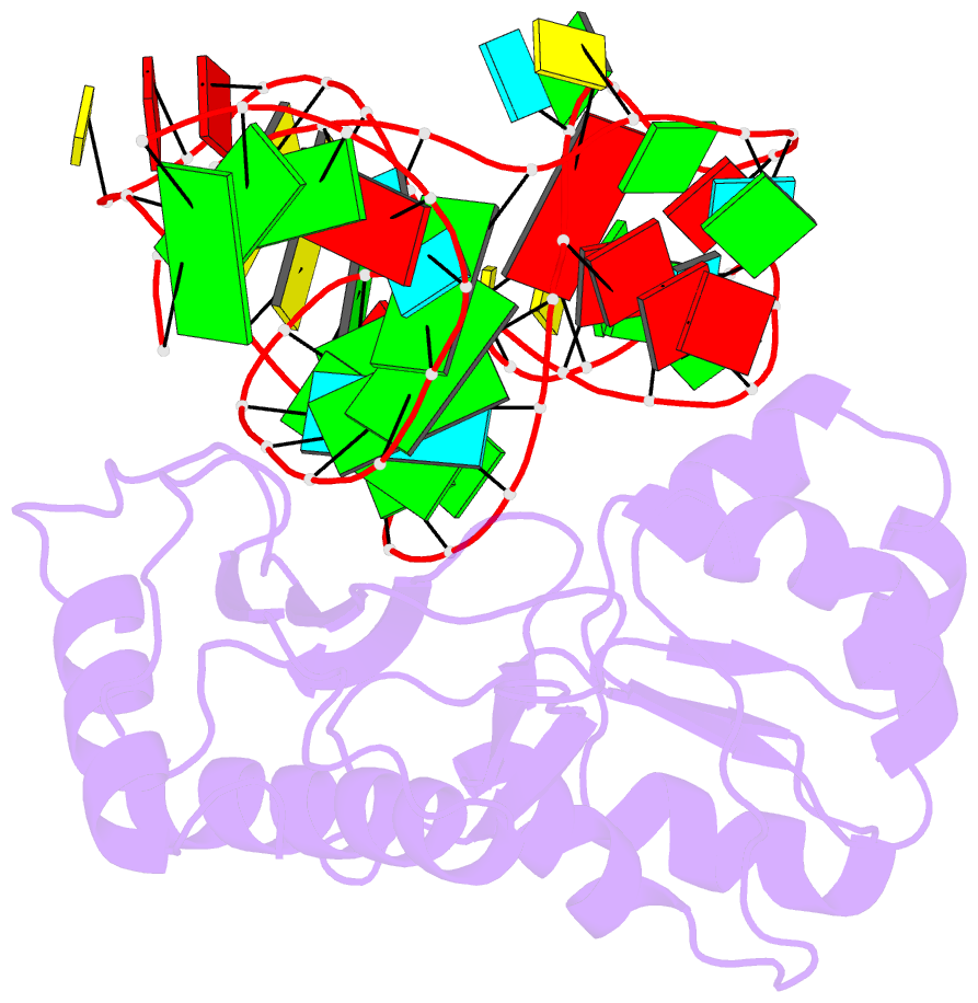
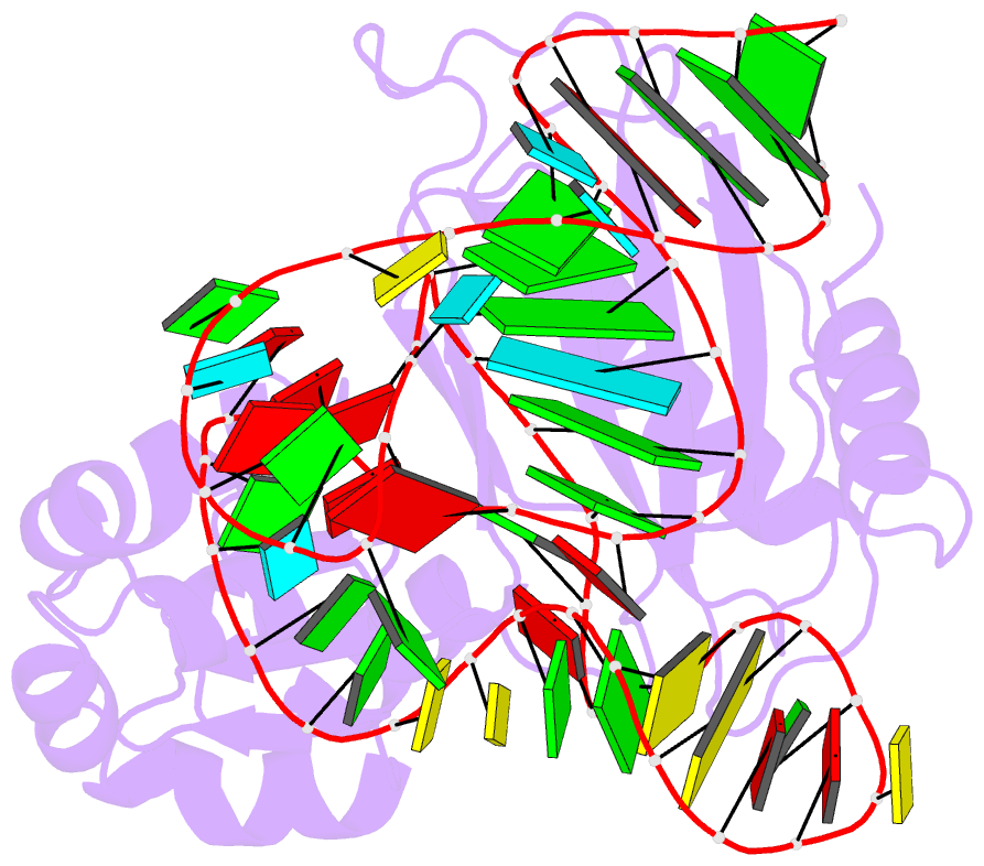
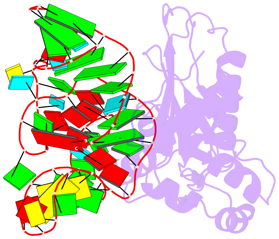
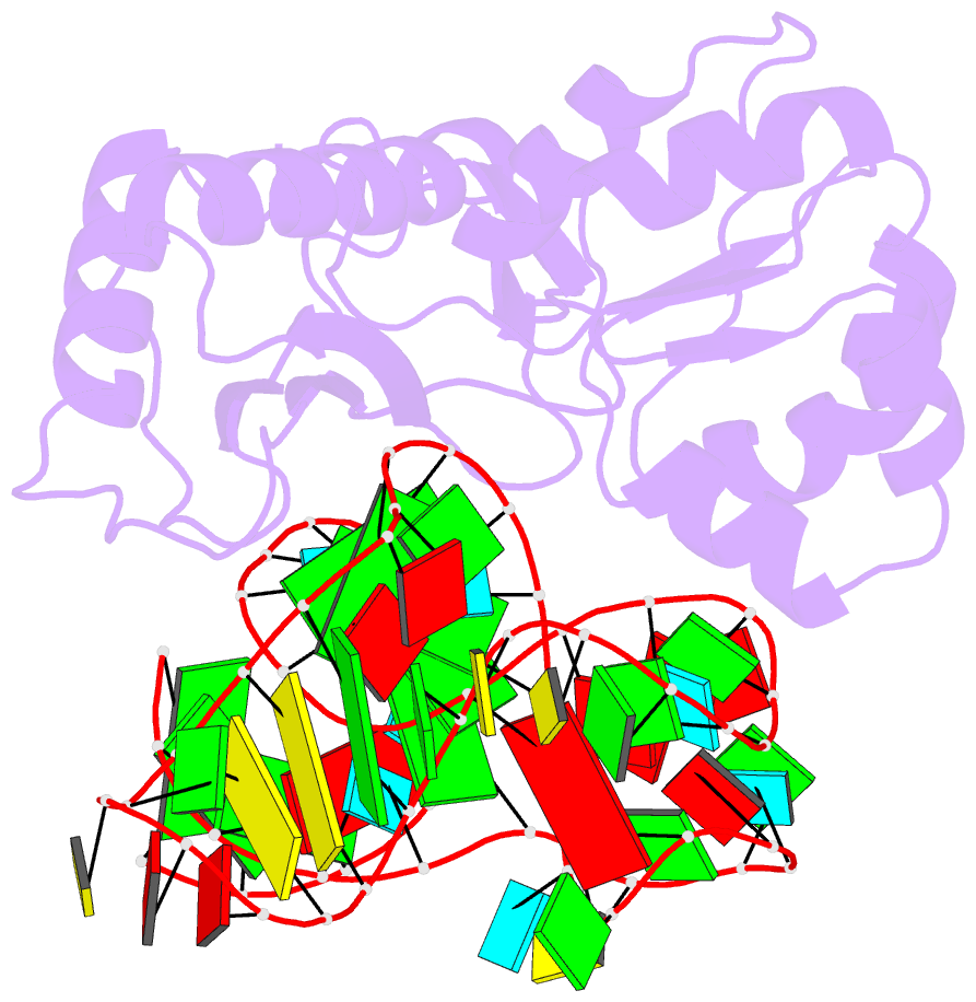
- Structure of the l1 protuberance in the ribosome
- Reference
- Nikulin A, Eliseikina I, Tishchenko S, Nevskaya N, Davydova N, Platonova O, Piendl W, Selmer M, Liljas A, Drygin D, Zimmermann R, Garber M, Nikonov S (2003): "Structure of the L1 protuberance in the ribosome." Nat.Struct.Biol., 10, 104-108. doi: 10.1038/nsb886.
- Abstract
- The L1 protuberance of the 50S ribosomal subunit is implicated in the release/disposal of deacylated tRNA from the E site. The apparent mobility of this ribosomal region has thus far prevented an accurate determination of its three-dimensional structure within either the 50S subunit or the 70S ribosome. Here we report the crystal structure at 2.65 A resolution of ribosomal protein L1 from Sulfolobus acidocaldarius in complex with a specific 55-nucleotide fragment of 23S rRNA from Thermus thermophilus. This structure fills a major gap in current models of the 50S ribosomal subunit. The conformations of L1 and of the rRNA fragment differ dramatically from those within the crystallographic model of the T. thermophilus 70S ribosome. Incorporation of the L1-rRNA complex into the structural models of the T. thermophilus 70S ribosome and the Deinococcus radiodurans 50S subunit gives a reliable representation of most of the L1 protuberance within the ribosome.