Summary information and primary citation
- PDB-id
- 1oct; SNAP-derived features in text and JSON formats;
DNAproDB
- Class
- transcription-DNA
- Method
- X-ray (3.0 Å)
- Summary
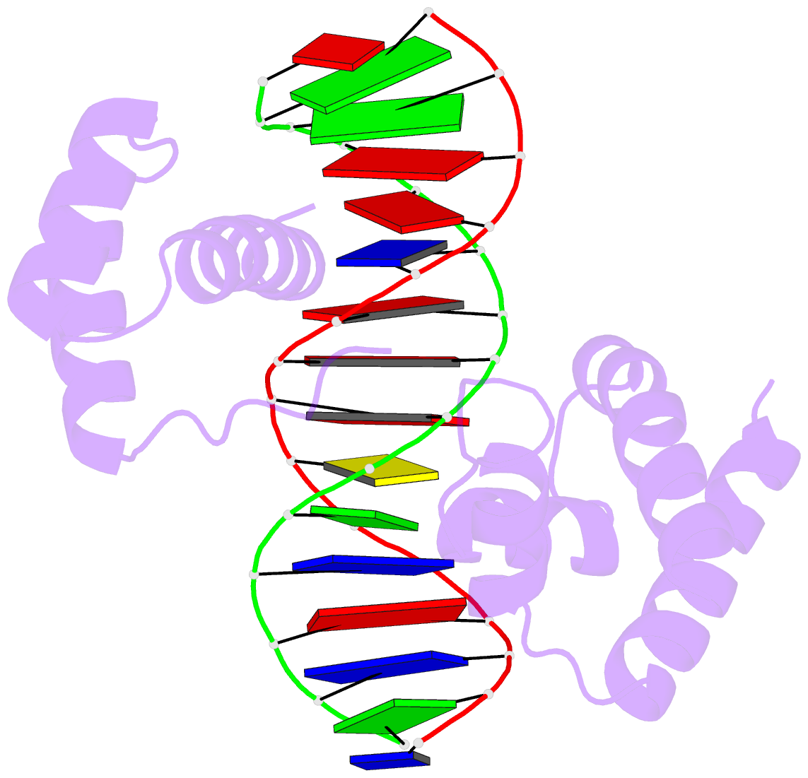
- Crystal structure of the oct-1 pou domain bound to an octamer site: DNA recognition with tethered DNA-binding modules
- Reference
- Klemm JD, Rould MA, Aurora R, Herr W, Pabo CO (1994): "Crystal structure of the Oct-1 POU domain bound to an octamer site: DNA recognition with tethered DNA-binding modules." Cell(Cambridge,Mass.), 77, 21-32. doi: 10.1016/0092-8674(94)90231-3.
- Abstract
- The structure of an Oct-1 POU domain-octamer DNA complex has been solved at 3.0 A resolution. The POU-specific domain contacts the 5' half of this site (ATGCAAAT), and as predicted from nuclear magnetic resonance studies, the structure, docking, and contacts are remarkably similar to those of the lambda and 434 repressors. The POU homeodomain contacts the 3' half of this site (ATGCAAAT), and the docking is similar to that of the engrailed, MAT alpha 2, and Antennapedia homeodomains. The linker region is not visible and there are no protein-protein contacts between the domains, but overlapping phosphate contacts near the center of the octamer site may favor cooperative binding. This novel arrangement raises important questions about cooperativity in protein-DNA recognition.


