Summary information and primary citation
- PDB-id
- 1odh; SNAP-derived features in text and JSON formats;
DNAproDB
- Class
- transcription factor-DNA
- Method
- X-ray (2.85 Å)
- Summary
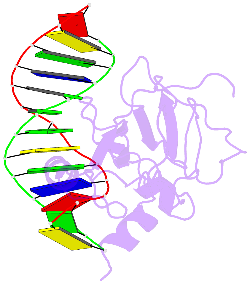
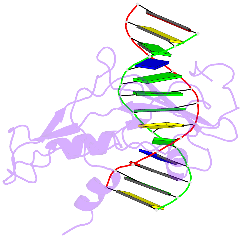
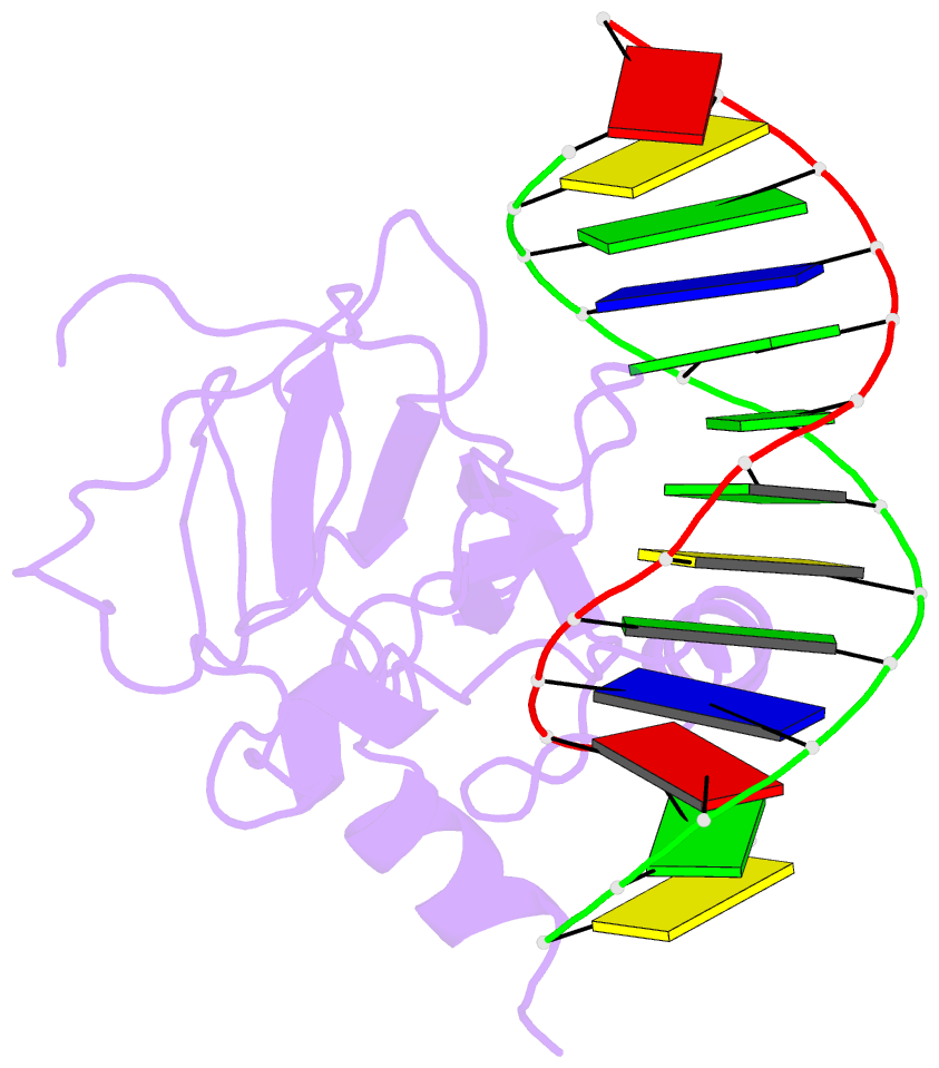
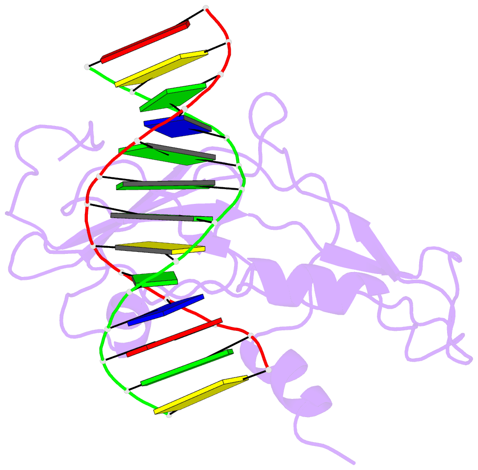
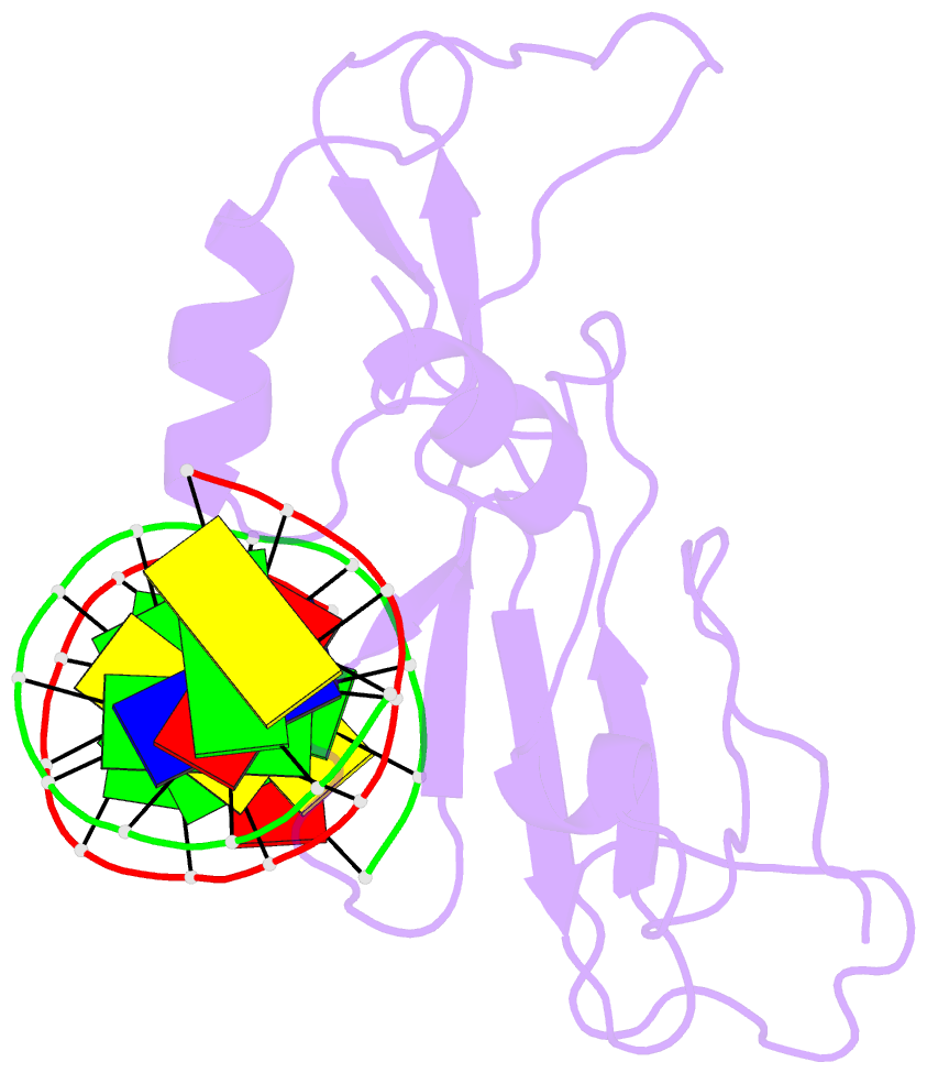
- Structure of the gcm domain bound to DNA
- Reference
- Cohen SX, Moulin M, Hashemolhosseini S, Kilian K, Wegner M, Muller CW (2003): "Crystal Structure of the Gcm Domain-DNA Complex: A DNA-Binding Domain with a Novel Fold and Mode of Target Site Recognition." Embo J., 22, 1835. doi: 10.1093/EMBOJ/CDG182.
- Abstract
- Glia cell missing (GCM) transcription factors form a small family of transcriptional regulators in metazoans. The prototypical Drosophila GCM protein directs the differentiation of neuron precursor cells into glia cells, whereas mammalian GCM proteins are involved in placenta and parathyroid development. GCM proteins share a highly conserved 150 amino acid residue region responsible for DNA binding, known as the GCM domain. Here we present the crystal structure of the GCM domain from murine GCMa bound to its octameric DNA target site at 2.85 A resolution. The GCM domain exhibits a novel fold consisting of two domains tethered together by one of two structural Zn ions. We observe the novel use of a beta-sheet in DNA recognition, whereby a five- stranded beta-sheet protrudes into the major groove perpendicular to the DNA axis. The structure combined with mutational analysis of the target site and of DNA-contacting residues provides insight into DNA recognition by this new type of Zn-containing DNA-binding domain.