Summary information and primary citation
- PDB-id
- 1p47; SNAP-derived features in text and JSON formats;
DNAproDB
- Class
- transcription-DNA
- Method
- X-ray (2.2 Å)
- Summary
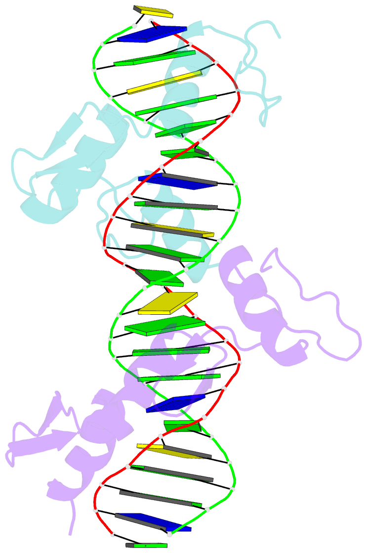
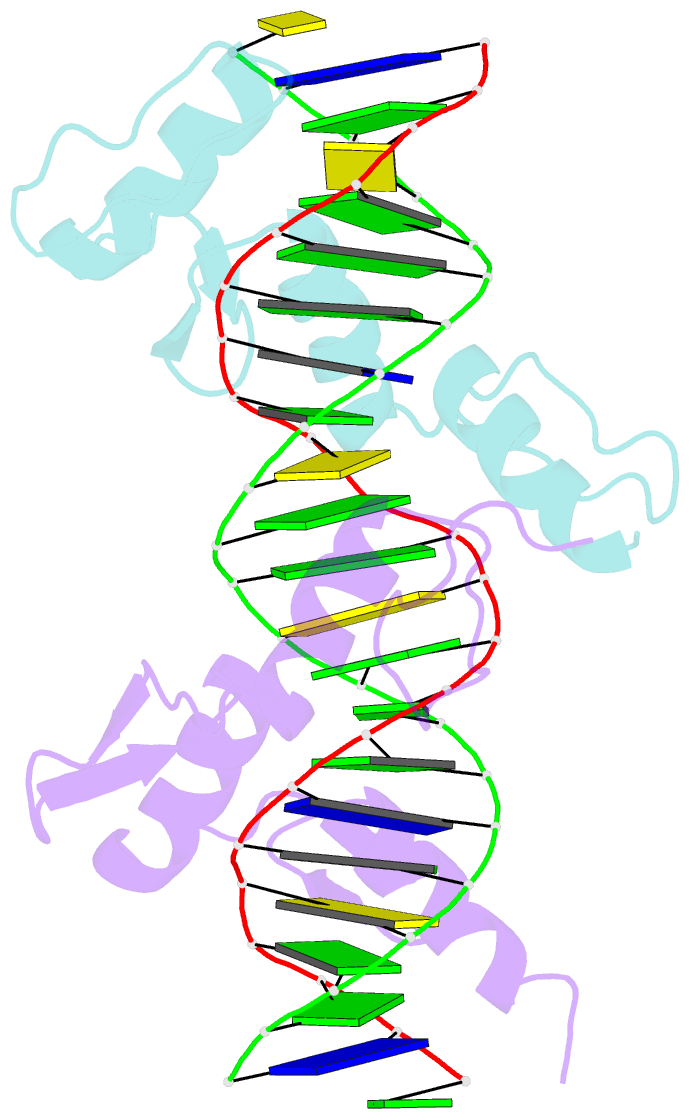
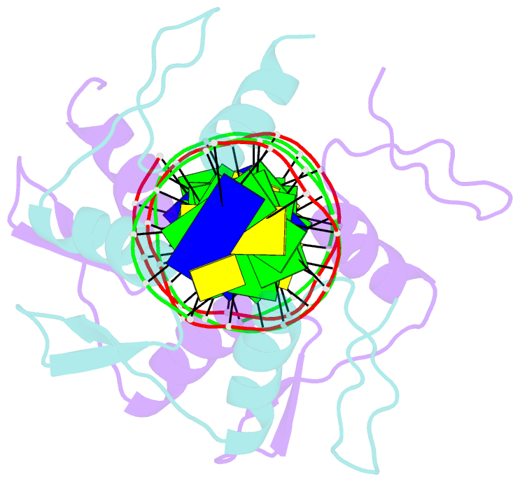
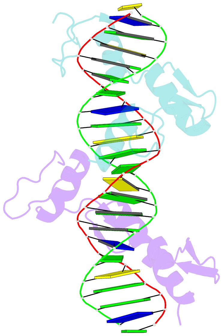
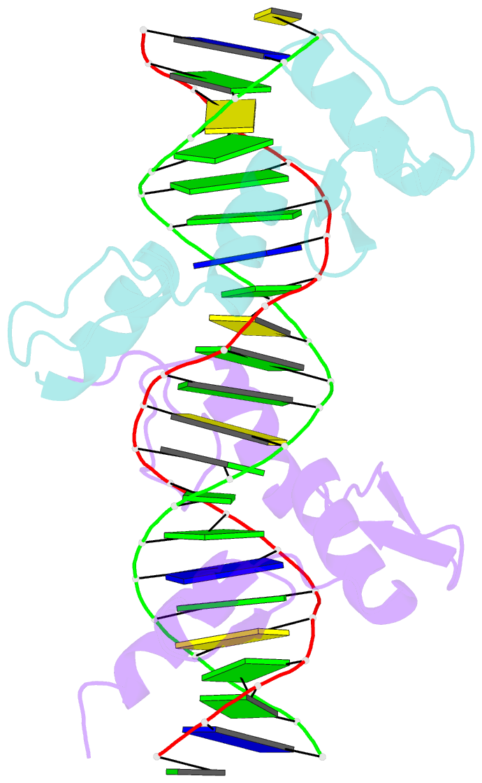
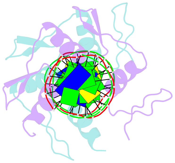
- Crystal structure of tandem zif268 molecules complexed to DNA
- Reference
- Peisach E, Pabo CO (2003): "Constraints for Zinc Finger Linker Design as Inferred from X-ray Crystal Structure of Tandem Zif268-DNA Complexes." J.Mol.Biol., 330, 1-7. doi: 10.1016/S0022-2836(03)00572-2.
- Abstract
- Zinc-finger proteins offer a versatile and effective framework for the recognition of DNA binding sites. By connecting multiple fingers together with canonical TGEKP linkers, a protein may be designed to recognize almost any desired target DNA sequence. However, proteins containing more than three zinc-fingers do not bind as tightly as one might predict, and it appears that some type of strain is introduced when a six-finger protein is constructed with canonical linkers. In an attempt to understand the sources of this strain, we have solved the 2.2A resolution X-ray crystallographic structure of a complex that has two copies of the three-finger Zif268 protein bound to adjacent sites on one duplex DNA. Conceptually, this is equivalent to a six-finger protein in which the central linker has been removed and the complex has been allowed to "relax" to its most stable conformation. As in other Zif268-DNA complexes, the DNA is approximately linear and is slightly underwound. Surprisingly, the structure of the complex is similar (within 0.5A) to an arrangement that would allow a canonical linker at the center of the complex, and it seems possible that entropic effects (involving the librational degrees of freedom in the complex) could be important in determining optimal linker length.