Summary information and primary citation
- PDB-id
- 1par; SNAP-derived features in text and JSON formats;
DNAproDB
- Class
- transcription-DNA
- Method
- X-ray (2.6 Å)
- Summary
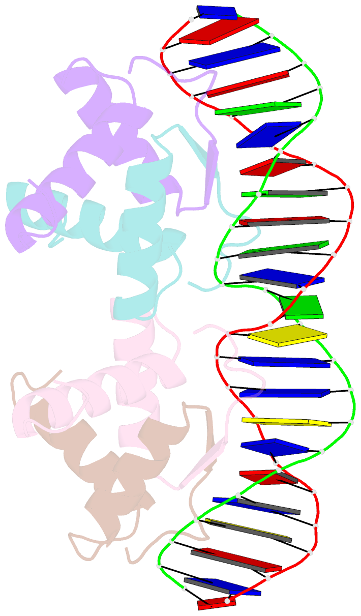
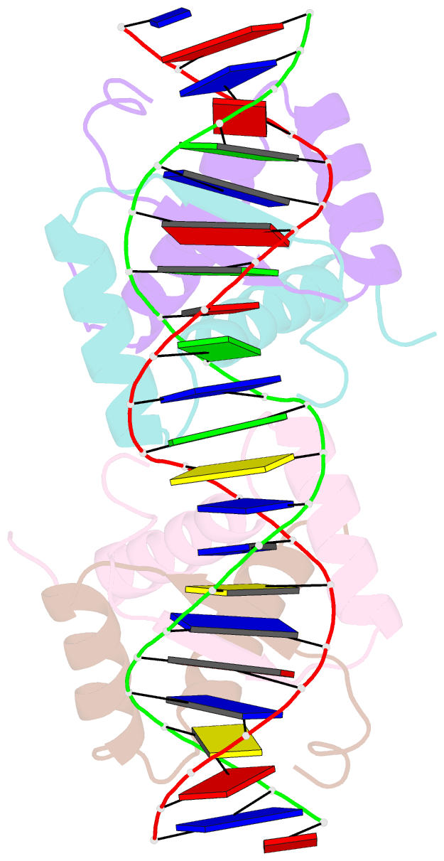
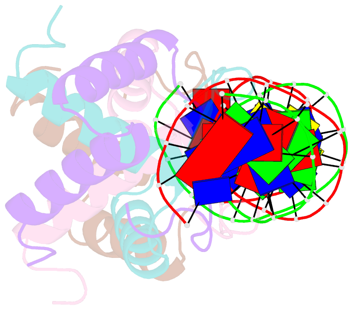
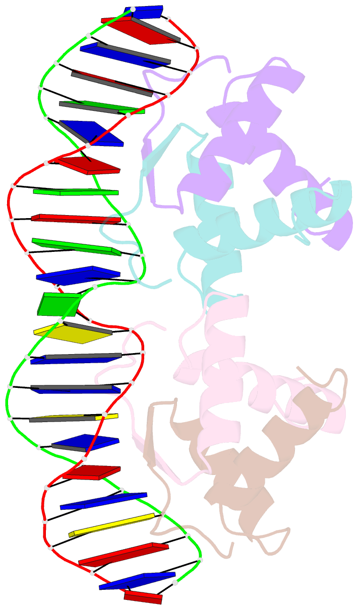
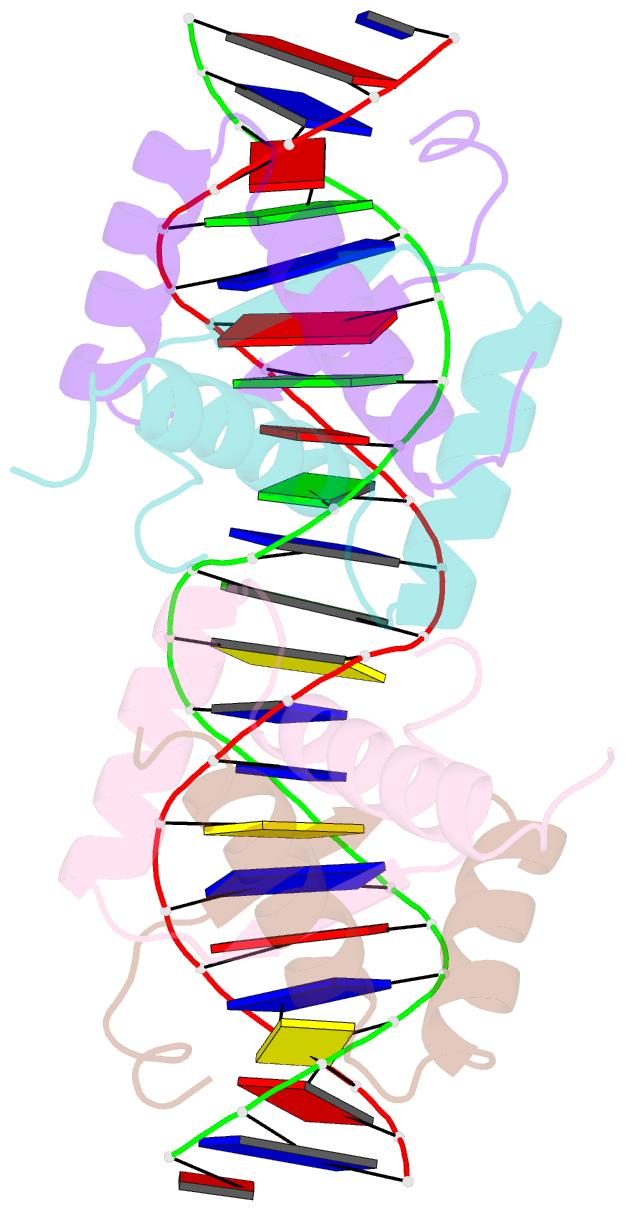
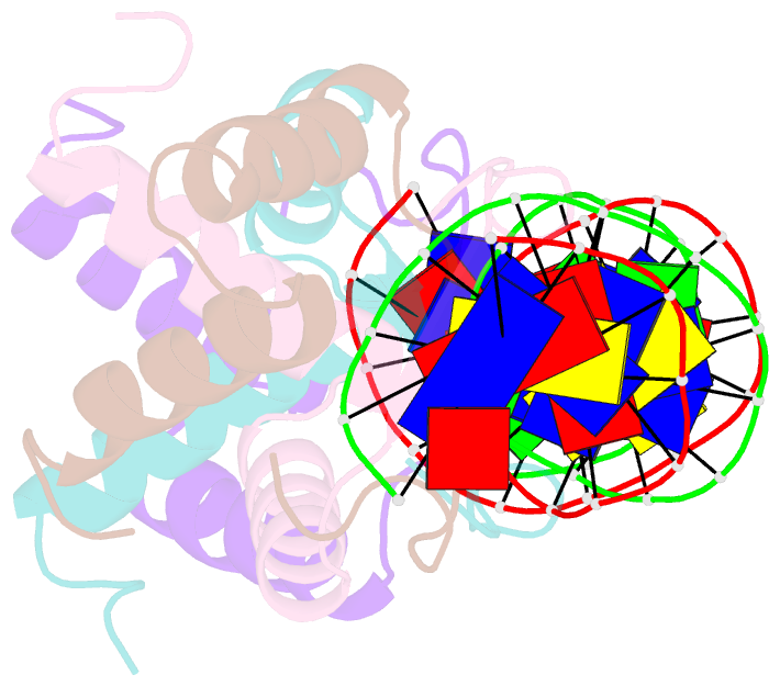
- DNA recognition by beta-sheets in the arc repressor-operator crystal structure
- Reference
- Raumann BE, Rould MA, Pabo CO, Sauer RT (1994): "DNA recognition by beta-sheets in the Arc repressor-operator crystal structure." Nature, 367, 754-757. doi: 10.1038/367754a0.
- Abstract
- Transcription of the ant gene during lytic growth of bacteriophage P22 (ref. 1) is regulated by the cooperative binding of two Arc repressor dimers to a 21-base-pair operator site. Here we report the co-crystal structure of this Arc tetramer-operator complex at 2.6 A resolution. As expected from genetic and structural studies and from the co-crystal structure of the homologous Escherichia coli MetJ repressor, each Arc dimer uses an antiparallel beta-sheet to recognize bases in the major groove. However, the Arc and MetJ complexes differ in several important ways: the beta-sheet-DNA interactions of Arc are far less symmetrical; DNA binding by Arc is accompanied by important conformational changes in the beta-sheet; and Arc uses a different part of its protein surface for dimer-dimer interactions.